More Recent Comments

Showing posts with label Nobel Prize. Show all posts

Thursday, August 13, 2009

Nobel Laureates: Peter Doherty and Rolf Zinkernagel


The Nobel Prize in Physiology or Medicine 1966

"for their discoveries concerning the specificity of the cell mediated immune defence"


Peter C. Doherty (1949 - ) and Rolf M. Zinkernagel (1940 - ) won the Noble Prize in 1996 for their work on the mechanism of cellular immunity (cell-mediated immunity). They were the ones who figured out how T-cells can recognize and kill cells that are infected with a virus.

This is part of the modern era of Nobel Prize awards where the Nobel Foundation is doing a wonderful job of explaining the awards to a scientifically literate general public. Here's the 1996 Press ReleaseTHEME:
Nobel Laureates
Summary

Peter Doherty and Rolf Zinkernagel have been awarded this year's Nobel Prize in Physiology or Medicine for the discovery of how the immune system recognizes virus infected cells. Their discovery has, in its turn, laid a foundation for an understanding of general mechanisms used by the cellular immune system to recognize both foreign microorganisms and self molecules. This discovery is therefore highly relevant to clinical medicine. It relates both to efforts to strengthen the immune response against invading microorganisms and certain forms of cancer, and to efforts to diminish the effects of autoimmune reactions in inflammatory diseases, such as rheumatic conditions, multiple sclerosis and diabetes.

The two Nobel Laureates carried out the research for which they have now been awarded the Prize in 1973-75 at the John Curtin School of Medical Research in Canberra, Australia, where Peter Doherty already held his position and to which Rolf Zinkernagel came from Switzerland as a research fellow. During their studies of the response of mice to viruses, they found that white blood cells (lymphocytes) must recognize both the virus and certain self molecules - the so-called major histocompatibility antigens - in order to kill the virus-infected cells. This principle of simultaneous recognition of both self and foreign molecules has since then constituted a foundation for the further understanding of the specificity of the cellular immune system.

The background to the Laureates' research

The immune system consists of different kinds of white blood cells, including T- and B- lymphocytes whose common function is to protect the individual against infections by means of eliminating invading microorganisms and infected cells. At the same time they must avoid damaging the own organism. What is required is a well developed recognition system that enables lymphocytes to distinguish between on the one hand microorganisms and infected cells, and on the other, the individual´s normal cells. In addition, the recognition system must be able to determine when white blood cells with a capacity to kill should be activated.

In the early 1970s when Peter Doherty and Rolf Zinkernagel had begun their scientific work within immunology, it was possible to distinguish between antibody-mediated and cell- mediated immunity. It was known that antibodies that are produced by B-lymphocytes are able to recognize and eliminate certain microorganisms, particularly bacteria. Far less was known about recognition mechanisms in the cellular immune system, for instance in conjunction with the killing of virus-infected cells by T-lymphocytes. One area where cellular immunity had previously been studied in some detail was, however, transplantation biology. It was known that T-lymphocytes could kill cells from a foreign individual after recognition of certain molecules - the major histocompatibility antigens - in the transplant.

The discovery

Rolf Zinkernagel and Peter Doherty used mice to study how the immune system, and particularly T -lymphocytes, could protect animals against infection from a virus able to cause meningitis. Infected mice developed killer T-lymphocytes, which in a test-tube could kill virus- infected cells. But there was an unexpected discovery: the T-lymphocytes, even though they were reactive against that very virus, were not able to kill virus-infected cells from another strain of mice. What decided whether or not a cell was eliminated by these killer lymphocytes was not only if they were infected with the virus, but also if they carried the "correct" variant of histocompatibility antigens, those of the infected mouse itself. Zinkernagel's and Doherty's findings, which were published in Nature in 1974 (1,2), demonstrated conclusively the requirement for the cellular immune system to recognize simultaneously both 'foreign' molecules (in the present case from a virus) and self molecules (major histocompatibility antigens). What also became obvious was the important function of the major histocompatibility antigens (in man called HLA-antigens) in the individual´s normal immune response and not only in conjunction with transplantation.

The discovery has given an impetus to later research

Zinkernagel's and Doherty´s findings had an immediate impact on immunological research. The wide relevance of their observations concerning the specificity of the T-lymphocytes became apparent in many contexts, both in regard to the ability of the immune system to recognize microorganisms other than viruses, and in regard to the ability of the immune system to react against certain kinds of self tissue. To explain their findings, the two scientists subsequently devised two models; one model was based on a single recognition of 'altered self''(when the histocompatibility antigen has been modified through association with a virus), the other on a 'dual recognition' of both foreign and self. (Fig.) Both the experimental findings and the theoretical models became immensely important in later research. Within a few years, it had been demonstrated that the set of the T- lymphocytes that are allowed to mature and survive in an individual is determined by the ability of the cell to recognize the transplantation antigens of the individual. Therefore, the principle of simultaneous recognition is essential for the ability of the immune system to distinguish between 'self' and 'non-self'.

Further molecular research has both confirmed Zinkernagel's and Doherty's models and clarified the structural basis of their discovery - that a small part (a peptide), for example from a virus, is directly bound to a defined variable part of the body´s own histocompatibility antigens, and that this complex is what is recognized by the specific recognition molecules of T- lymphocytes (T-cell receptors). Taken in all, the clarification of the recognition mechanisms of the T-cells within the cellular immune system has fundamentally changed our understanding of the development and normal function of the immune system and, in addition, has also provided new possibilities for the selective modification of immune reactions both to microorganisms, and to self tissues.

Figure legend: The figure describes how a killer T lymphocyte must recognize both the virus antigen and the self histocompatibility antigen molecule in order to kill a virus-infected target cell. The figure is a modification of the figure published by Zinkernagel and Doherty already 1974 (in Nature 251, p 547).

Relevance for clinical medicine

Many common and severe diseases depend on the function of the cellular immune system and consequently on its mechanisms for specific recognition. Although this naturally applies to infectious diseases, this is also true of a number of chronic inflammatory conditions such as rheumatic diseases, diabetes and multiple sclerosis. Where infectious diseases are concerned, the new knowledge provides a better platform for the construction of new vaccines; one can ascertain exactly what parts of a microorganism are recognized by the cellular immune system, and can specifically focus the production of the vaccine on those parts. Furthermore, regard is paid to the fundamental principles formulated by Doherty and Zinkernagel in trials with vaccination against the emergence of metastases in certain forms of cancer. In many chronic inflammatory diseases, better explanations have been provided for the associations between disease susceptibility and the histocompatibility antigen type carried by an individual. The research that followed from the now awarded discovery has also provided openings for selectively diminishing or altering immune reactions that play a central role in inflammatory diseases.

References

1. Zinkernagel RM, Doherty PC. Restriction of in vitro T cell-mediated cytotoxicity in lymphocytic choriomeningitis within a syngenic and semiallogeneic system. Nature 248, 701- 702, 1974.

2. Zinkernagel RM, Doherty PC. Immunological surveillance against altered self components by sensitised T lymphocytes in lymphocytic choriomeningitis. Nature 251, 547-548, 1974.

3. Doherty PC, Zinkernagel RM. A biological role for the major histocompatibility antigens. Lancet, 1406-1409, 1975.

4. Zinkernagel RM, Doherty PC. MHC restricted cytotoxic T cells: Studies on the biological role of polymorphic major transplantation antigens determining T cell restriction specificity. Advances in Immunology 27, 51-177, 1979.


The images of the Nobel Prize medals are registered trademarks of the Nobel Foundation (© The Nobel Foundation). They are used here, with permission, for educational purposes only.

Wednesday, July 29, 2009

Nobel Laureate: Roger Tsien


The Nobel Prize in Chemistry 2008.

"for the discovery and development of the green fluorescent protein, GFP"


Roger Y. Tsien (1952 - ) won the Nobel Prize in Chemistry for his work on adapting green fluorescent protein to serve as a marker for detecting changes within a cell. He shared the prize with Osamu Shimomura and Martin Chalfie.

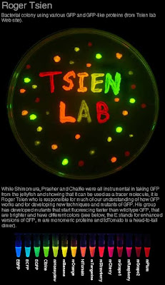
Tsien's lab learned what causes the protein to flouresce and how to modify the active site on order to create variants that emitted different colors. You can watch an excellent video of his Nobel Lecture: Constructing and Exploiting the Fluorescent Protein Paintbox.

If that's a little too technical, the work is described in a special document called Information for the Public.
THEME:
Nobel Laureates
This is where the third Nobel Prize laureate Roger Tsien makes his entry. His greatest contribution to the GFP revolution was that he extended the researchers’ palette with many new colours that glowed longer and with higher intensity.

To begin with, Tsien charted how the GFP chromophore is formed chemically in the 238-aminoacid-long GFP protein. Researchers had previously shown that three amino acids in position 65–67 react chemically with each other to form the chromosphore. Tsien showed that this chemical reaction requires oxygen and explained how it can happen without the help of other proteins.

With the aid of DNA technology, Tsien took the next step and exchanged various amino acids in different parts of GFP. This led to the protein both absorbing and emitting light in other parts of the spectrum. By experimenting with the amino acid composition, Tsien was able to develop new variants of GFP that shine more strongly and in quite different colours such as cyan, blue and yellow. That is how researchers today can mark different proteins in different colours to see their interactions.

One colour, however, that Tsien could not produce with GFP was red. Red light penetrates biological tissue more easily and is therefore especially useful for researchers who want to study cells and organs inside the body.

At this point, Mikhail Matz and Sergei Lukyanov, two Russian researchers, became involved in the GFP revolution. They looked for GFP-like proteins in fluorescent corals and found six more proteins, one red, one blue and the rest green.

The desired red protein, DsRED, was unfortunately larger and heavier than GFP. DsRED consisted of four amino acid chains instead of one and was of less use as a fluorescent tag in biological processes. Tsien’s research group solved this problem, redesigning DsRED so that the protein is now stable and fluoresces as a single amino acid chain, which can easily be connected to other proteins.

From this smaller protein, Tsien’s research group also developed proteins with mouth watering names like mPlum, mCherry, mStrawberry, mOrange and mCitrine, according to the colour they glowed. Several other researchers and companies have also contributed new colours to this growing palette. So today, 46 years after Shimomura first wrote about the green fluorescent protein, there is a kaleidoscope of GFP-like proteins which shine with all the colours of the rainbow.

The brainbow

Three of these proteins have been used by researchers in a spectacular experiment. Mice were genetically modified to produce varying amounts of the colours yellow, cyan and red within the nerve cells of their brain. This combination of colours is similar to the one used by computer printers. The result was a mouse brain that glowed in the colours of the rainbow. The researchers could follow nerve fibres from individual cells in the dense network in the brain.

The researchers called this experiment “the brainbow”.


[Photo Credit: UCSD]

The images of the Nobel Prize medals are registered trademarks of the Nobel Foundation (© The Nobel Foundation). They are used here, with permission, for educational purposes only.

Friday, July 24, 2009

Nobel Laureate: Peyton Rous


The Nobel Prize in Physiology or Medicine 1966

"for his discovery of tumour-inducing viruses"


Peyton Rous (1879 - 1970) won the Noble Prize in 1966 for his discovery and characterization of several viruses that cause cancer in birds and mammals. He is best known for his work on Rous Sarcom Virus (RSV), a retrovirus that infects chickens and causes cancer.

The original work was done many years earlier as he describes in his Nobel lecture.
In 1910 I described a malignant chicken sarcoma which could be propagated by transplanting its cells, these multiplying in their new hosts and forming new tumors of the same sort. In other ways the growth showed itself to be a neoplasm of a classical sort, yet, as reported in 1911, its cells yielded a causative virus. Numerous workers had already tried by then to get extraneous causes from transplanted mouse and rat tumors but the transferred cells had held their secret close. Hence the findings with the sarcoma were met with down-right disbelief, though soon several other, morphologically different, "spontaneous" chicken tumors were propagated by transplantation and from each a virus was got causing growths of its kind. Not until after some 15 years of disputation amongst oncologists were the findings with chickens deemed valid, and then they were relegated to a category distinct from that of mammals because from them no viruses could be obtained. Only in 1925, through the efforts of a British worker, W.E. Gye, was much attention given them by scientists.

The virus causing the chicken sarcoma first studied, now generally termed the RSV, has been maintained for more than fifty-five years and is still studied in many countries. Throughout most of this time it would engender growths only in chickens and closely related fowls; but of late several extraneous, non-neoplastic viruses have become associated with it, during its passage in unusual avian hosts; and its scope has thus been so enlarged that now not at few mammals, including monkeys, have been found to develop tumors after inoculation with the enhanced material
For many years the idea that a virus could cause cancer was not part of mainstream oncology. It took a number of other advances to make the idea acceptable. Part of the problem was due to a lack of understanding about the various modes of viral infection. Work on bacterial viruses (bacteriophage) showed clearly that a virus genome could integrate into the normal cellular chromosome and be carried along in all the offspring of those cells.

Fifty years after the initial discovery of a cancer-causing virus, scientists could finally begin to formulate a mechanism based on the results of the phage ground and bacterial geneticists. It's interesting to see the state of knowledge in 1966 as seen in the presentation speech.
THEME:
Nobel Laureates
The situation changed radically in the 1950's. The study of tumour viruses is a central area of modern cancer research. Two developments are responsible for this remarkable change. Recent developments in microbial genetics have lead to reinterpretation of the virus concept itself. It turns out that certain types of virus can introduce parts of their own genetic material into a cell without killing it or inhibiting its multiplication. The virus material thus introduced may become actually integrated with the gene material of the recipient cell and behave as a new hereditary factor. Virus infection can thus lead to a permanent change in some cellular characteristics. This re-evaluation of the virus concept made it possible to understand how a tumour virus might change the regulated behaviour of normal cells to the malignant proliferation characteristic of cancer cells. In the same period many new viruses capable of inducing malignant tumours in mammals were discovered. In 1981 Gross found a virus that induces leukemia in mice. A few years later he and two women scientists, Stewart and Eddy, isolated a remarkable new virus, called polyoma, capable of inducing an array of tumours in many different mammalian species. Since 1960 more than a dozen new tumour virus types have been isolated. It was established, furthermore, that tumour viruses could change normal to cancer cells in the test tube after a very short time of contact. This opened the way for direct studies on cancerous transformation of human cells, an approach previously hidden behind the walls of the living organism. Remarkably enough, it could be shown that Rous' own virus, previously regarded as lacking any importance for mammals, induces cancer under certain conditions in many different mammalian species and may even transform human cells in test tube cultures. Swedish scientists in Lund and Uppsala have made important contributions in this regard. It is not yet clear in which way viruses induce cancer but there is much to indicate that the virus does not behave like a little boy setting fire to a hayrick and running away; part of the viruses' own genetic material seems to be directly responsible for the malignant behaviour of the virus transformed tumour cell.


The images of the Nobel Prize medals are registered trademarks of the Nobel Foundation (© The Nobel Foundation). They are used here, with permission, for educational purposes only.
Monday, July 20, 2009

Nobel Laureates: J. Michael Bishop and Harold Varmus


The Nobel Prize in Physiology or Medicine 1989

"for their discovery of the cellular origin of retroviral oncogenes"


J. Michael Bishop (1936 - ) and Harold E. Varmus (1939 - ) won the Noble Prize in 1989 for proving that viruses contain a cancer-causing gene derived from the genome of the organism they infect. Specifically, they showed that chicken Rous Sarcoma Virus (RSV) carried an oncogene called v-src and this gene was an intronless version of a normal chicken gene called c-src.

The discovery had tremendous implications in many fields. Not only did it explain the origins of a particular chicken cancer but it also suggested that there where many other oncogenes in other well-known retroviruses. This turned out to be correct and several dozen such oncogenes have been discovered (e.g. abl, fos, jun, and erb among others).

The discovery also ignited a burst of activity in signaling because the c-src gene encodes a tyrosine kinase. This is an enzyme that adds phosphate groups to proteins thereby affecting their activity. Such enzymes belong to pathways triggered by specific signals leading to the regulation of growth and cell division.

The discovery lent support to the idea that small changes in regulatory pathways could have large effects—in this case converting a normal cell to a cancer cell. This idea fit nicely with the work of developmental biologists who were showing that single genes could regulate entire developmental pathways. It meant that large-scale changes in morphology during evolution could be effected by small changes (mutations) in the genome.

Finally, the work of Bishop and Varmus helped change our view of the genome. Not only do retrovirus sequences pop in and out of the genome but they can also capture cellular genes by converting them to retrogenes. The work confirmed the idea that genomes were dynamic—a idea that began with transposons. Not only that, the discovery of the retrogene, v-src, showed us that introns were almost certainly not an essential component of a gene.

In my opinion, this is one of the most significant scientific advances of the 2oth century. There aren't many achievements that really count as breakthroughs because either the work was done in many labs and came out piecemeal, or the result wasn't very significant. This is one achievement that truly was a big step forward in biology.

By 1989, the press releases from the Karolinska Institute were becoming excellent educational tools for the scientifically literate general public. In this case, the press release explains how oncogenes cause cancer. I'm going to include the entire Press Release (below) because it's so good.
THEME:
Nobel Laureates
Summary

The discovery awarded with this year's Nobel Prize in Physiology or Medicine concerns the identification of a large family of genes which control the normal growth and division of cells. Disturbances in one or some of these so-called oncogenes (Gk ónco(s) bulk, mass) can lead to transformation of a normal cell into a tumor cell and result in cancer.

Michael Bishop and Harold Varmus used an oncogenic retrovirus to identify the growth-controlling oncogenes in normal cells. In 1976 they published the remarkable conclusion that the oncogene in the virus did not represent a true viral gene but instead was a normal cellular gene, which the virus had acquired during replication in the host cell and thereafter carried along.

Bishop's and Varmus' discovery of the cellular origin of retroviral oncogenes has had an extensive influence on the development of our knowledge about mechanisms for tumor development. Until now more than 40 different oncogenes have been demonstrated. The discovery has also widened our insight into the complicated signal systems which govern the normal growth of cells.

Cellular Oncogenes Discovered by the Use of Retrovirus

The term oncogene was introduced in the middle of the 1960s to denote special parts of the genetic material of certain viruses. It was believed that this part of the genetic material could direct the transformation of a normal cell into a tumor cell under the influence of other parts of the viral genetic material, alternatively via chemical or physical effects. The favourite theory of the time was that virus-mediated cell-to-cell transmittance of oncogenes was the origin of all forms of cancer. This view was later proven to be incorrect.

The original discovery of an oncogenic virus was made in 1916 by Peyton Rous working at the Rockefeller Institute in New York. Fifty years later Rous received the Nobel Prize in Physiology or Medicine. Rous virus, as the infectious agent later was named, is a member of a large virus family named retroviruses. The genetic material of these viruses is RNA (ribonucleic acid). This RNA can be transcribed into DNA (deoxyribonucleic acid) by a unique enzyme in the virus, reverse transcriptase. The 1975 Nobel Prize in Physiology or Medicine was awarded to David Baltimore, Renato Dulbecco and Howard Temin partly for the discovery of this enzyme.

Reverse transcription of the genetic material of the virus into DNA has the important consequence that it can become integrated into the chromosomal DNA in the cells. It was through investigations of Rous virus that this year's laureates Michael Bishop and Harold Varmus in 1975 could demonstrate the true origin of oncogenes. They used one variant of Rous virus which contained an oncogenic gene (Figure 1) and another variant which lacked this gene. By use of these viruses they managed to construct a nucleic acid probe which selectively identified the oncogene. This probe was used to search for the corresponding genetic material in DNA from different cells. It was then found that oncogene-like material could be detected in different species throughout the animal kingdom, in fact even in simple organisms comprising only a few cells. Furthermore, it was shown that the gene had a fixed position in the chromosomes of a certain species, and that the gene, when it constituted part of the cellular genetic material, was divided into fragments (a mosaic gene) (Figure 1).
Figure 1. The difference between an oncogene in a virus and in a cell. In retroviruses causing tumors there is a separate segment of transforming nucleic acid which has been derived from a cell. The cellular gene is split (a mosaic gene) whereas the oncogene in the virus is continuous.
These findings led to the remarkable conclusion that the oncogene in the virus did not represent a true viral gene but a cellular gene which the virus had picked up far back during its replication in cells and carried along. This cellular gene was found to have a central function in the cells. It controlled their growth and division.

Through these studies of the abnormal, i.e. the diseased state, it was possible to elucidate critical normal cellular functions - a not uncommon situation in biomedical research. The original discovery of a cellular oncogene led to an intensive search for further similar genes. The explosive development of this field of research has led to the identification of more than 40 different oncogenes which direct different events in the complex signal systems that regulate the growth and division of cells. Changes in any one or more of these oncogenes may lead to cancer.

Balanced Cellular Interactions - A Biological Wonder

Symmetrical and asymmetrical, multicellular structures develop from the fertilized ovum by a process of differentiation about which only limited knowledge is available. In the fully developed individual carefully balanced conditions prevail. Damage of an organ elicits sophisticated repair processes which lead to restitution of the original condition of the organ. However, if a single cell escapes the network of growth control the result may be an abnormal local proliferation of cells or in the worst case a cancer implying the dissemination of cells running amok.

The development of a cancer is a complicated process involving several consecutive changes of the genetic material. Studies of cellular genes (proto-oncogenes) corresponding to the viral oncogenes, has started to shed light on the intricate systems which control normal cellular growth and division.

Cellular Oncogene Products Constitute Links in Signal Chains which Regulate Growth and Division of Cells

The regulation of growth and division of cells has turned out to be much more complicated than originally believed. Cellular oncogene products with different properties act in different positions of elaborate signal systems (Figure 2). In order to transmit signals from one cell to the other or from one cell to itself there are growth factors. These factors appear in the fluids surrounding cells. There are examples of oncogene products, viz. proteins produced in the cytoplasm, which can act as growth factors. Thus, it was found that the product of the sis1) gene was closely related to a previously identified growth factor PDGF (Platelet Derived Growth Factor).
Figure 2. Oncogene products are links in signal chains that stretch from the cell surface to the genetic material in the cell nucleus. This chain is composed of (1) growth factors, (2) growth factor receptors, (3) signal transducing proteins in cell membranes, (4) phosphokinases in the cytoplasm and (5) proteins transported from the cytoplasm into the nucleus where they bind to DNA. The localization of different oncogene products (Sis, ErbB, Ras, Src, Myc) is schematically indicated.
In order for a growth factor to be able to interact with a cell there has to be membrane structures, receptors, to which they can bind. There are several oncogene products which represent receptors in the cytoplasmic membrane of the cells, e.g. ErbA, Fms, Kit. These receptors have a unique enzymatic activity. They are so-called kinases with a capacity to phosphorylate (=add a phosphate group) the amino acid tyrosine. There are two more groups of oncogene products with phosphokinase activity; firstly tyrosine/phosphokinase which lack receptor function and is located at the inside of the cytoplasmic membrane, and secondly serine/threonine phosphokinase which is found in the cytoplasm.

Thus, oncogene products function as links in signal chains stretching from the surface of the cell to the genetic material in the nucleus. In the cytoplasm there is one more group of oncogene products. These are called Ras and are related to important cellular signal factors called G-proteins.

Finally, there is a large number of oncogene products which are located in the nucleus of the cell, i.e. Myc, Myb, Fos, ErbA and others. These products direct the transcription of DNA into RNA and therefore play a critical role in the selection of proteins to be synthesized by the cell.

Cancer - A Complex, Biological Sequence of Events

Changes in the genetic material constitute the basis for the development of all cancer. Generally there are several consecutive such changes which influence different steps in the signal chains described above. Therefore, one should à priori not expect to find one single clue to the mechanism of origin of cancer. However, application of the expanding knowledge in the oncogene field allows us to start comprehending the disharmonic orchestration behind abnormal cellular growth.

It is conceptually incorrect to speak about "cancer genes". However, historical circumstances explain why the oncogene terminology was introduced before a designation of the corresponding normal cellular genes was proposed. From the point of view of cancer the important matter is to compare oncogenes in normal cells and in tumor cells.

Oncogenes as a Cause of Cancer

The majority of oncogenes have been discovered in experimental studies using retroviruses. However, in a few cases oncogenes were identified by the use of an alternative technique, i.e. genetic material was isolated from tumor cells of non-viral origin and transferred (transfected) to other cells prapagated in culture. The cells receiving the DNA changed growth pattern, and further characterization of the transfected genetic material revealed the presence of oncogenes.

Two principally different forms of activation of oncogenes can be distinguished. Firstly, the normal cellular oncogene is hyperactive, and secondly the oncogene product is altered so that it can no longer be regulated in a normal way. There are several examples of these types of activation of oncogenes.

The discovery of oncogenes was as mentioned originally made by the use of retroviruses. This infers that genetic control elements in the virus itself can be responsible for the abnormal expression of the oncogene. However, in many cases it was found that alterations of the transferred oncogene contributed to its accentuated expression.

There are retroviruses which lack oncogenes but still can induce cancer. This is due to the fact that the virus has inserted its genetic material (in the form of DNA) very close to a normally occurring oncogene in the genetic material of the cell. This may result in an increased turn-over of the oncogene which may lead to abnormal cellular growth. The corresponding phenomenon can also occur in the absence of retroviruses. In this case there is a reorganization of the genetic material in the cell. Such a reorganization may occur within a single chromosome or by exchange of material between chromosomes. Repeated copying of a normal oncogene can lead to its amplification in the chromosome and consequently to increased amounts of the oncogene product. In certain brain tumors, glioblastomas, an amplified erbB-gene has been found, and a correspondingly increased neu-gene activity was shown in some forms of breast cancer.

The same effect can be seen when there is a reciprocal exchange of segments between chromosomes (translocation). Thus the normal myc-gene on chromosome 8 has been translocated to chromosome 14 in many patients with Burkitt's lymphomas (Figure 3). The insertion of the myc-gene containing chromosome segment is such that it becomes located close to hyperactive genes directing the synthesis of antibody protein. As a consequence the myc-gene becomes activated. Chromosome translocations occur in many different tumors. Chromosome analysis can therefore be of considerable value for localization of genetic changes in the genome critical for tumor development.
Figure 3. Chromosome translocation in Burkitt's lymphoma. Segments have been exchanged between chromosomes 8 and 14 which has activated the oncogene myc.
Oncogenes with point mutations have been observed in many tumors. These mutations may cause alterations in the amino acid composition of the gene product. A well-known example of such a modification is the exchange of amino acid 12 from glycine to valine in the ras gene product which has been observed in human tumor material. The mutation may also be somewhat more extensive leading to the absence of part of the protein (deletion). Different examples of modified oncogenes in human tumor material are given in Table I.
The Importance of Viruses for Cancer in Man

Cancer is not a contagious disease. However, infectious agents like viruses can contribute to the origin of cancer. Thus, it is by use of retroviruses that most oncogenes were identified, the starting materials in such investigations often being highly specialized, experimentally derived tumors. It seems likely that retroviruses play a relatively limited role for the development of cancer under natural conditions. The only known example in man in which a retrovirus infection contributes to the origin of cancer is the HTLV-1 associated lymphomas which occur in Japan.

However, there are other kinds of viruses which can contribute to the development of tumors in man. All these viruses have DNA as their genetic material. As examples can be mentioned papillome (wart) viruses and Epstein-Barr virus, a type of herpes virus. Certain types of papillome viruses play a role for the development of cervical cancer in the genital tract, while Epstein-Barr virus is an important factor for the development of Burkitt's lymphomas in Africa and nasopharyngeal cancer in Asia. However, in all these cases factors in addition to the virus infections are required for the cancer to develop.

References

J.M. Bishop: Oncogenes. Scientific American, 1982, 246, 68-78.

T. Hunter: The Proteins of Oncogenes. Scientific American, 1984, 251, 60-69.

C-H. Heldin & B. Westermark: Tillväxtfaktorer och onkgener. Läkartidningen 1988, 85, 497-499.

E. Norrby: I: Våra virus. Virus och cancer. Allmänna Förlaget, 1987, sid. 66-74.

1) All oncogenes are identified by the use of three letter abbreviations. In addition cellular and viral oncogenes are sometimes distinguished by c- and v- prefixes, respectively, e.g. c-src and v-src.


[Photo Credit: UCSF]

The images of the Nobel Prize medals are registered trademarks of the Nobel Foundation (© The Nobel Foundation). They are used here, with permission, for educational purposes only.

Friday, July 10, 2009

Nobel Laureate: Adolf Butenandt


The Nobel Prize in Chemistry 1939.

"for his work on sex hormones"


Adolf Friedrich Johann Butenandt (1903 - 1995) won the Nobel Prize in Chemistry for his work on the structure and function of sex hormones, particularly estradiol, progesterone, and testosterone.

He was not allowed to accept the Nobel Prize in 1939. After the war, in 1949, he was given the medal at a special ceremony.

He shared the 1939 Nobel Prize with Leopold Ruzicka.

The original (1939) award presentation describes Butenandt's isolation and identification of estradiol.
THEME:
Nobel Laureates
As recently as twelve years ago, very little was known about the nature of the sex hormones. As regards the oestrogenic, or follicle, hormone it was established that extracts from certain organs, e.g. the ovaries and placenta, bring about the characteristic oestrus phenomena in castrated female rats. Only a few observations were available concerning the stability and solubility of their active principles. Further development in the chemistry of the oestrogenic hormones could not take place until the purely biological discoveries by Allen and Doisy in 1923 and by Aschheim and Zondek in 1927 had been made.

Butenandt made the first big step forward in clarifying the chemistry of the follicle hormone in 1929 in Göttingen, simultaneously with Doisy in the United States. Both workers succeeded in isolating from the urine of pregnant women a substance in crystalline form having oestrogenic effects. Butenandt named this substance folliculine, a designation which was later changed to oestrone. He established that its empirical formula was C18H22O2, and that it was an oxyketone.

Shortly after the discovery of oestrone, Marrian in London (1930) isolated from the urine of pregnant women a new hormone which he called oestriol. Butenandt confirmed Marrian's discovery and explained the relationship between the new substance and oestrone. The relation between sterols and oestrogenic substances which had been assumed on crystallographical grounds became probable from the chemical point of view only after Butenandt and Marrian had shown, independently of one another, that only three benzoide double bonds enter into the ring system of these substances.

In 1932, Butenandt was able, from observations made in spectral analysis, and especially on the basis of the then established correct formula of cholesterol to draw up the formulae of the chemical structure of oestrone and oestriol. But there remained the important task of proving the chemical structure of the ring system as assumed by Butenandt. By breaking down the oestriol molecule stage by stage Butenandt proved that both œstrogenic hormones contained a phenanthrene core. At the same time he was able to obtain the same dimethyl phenanthrene from etiobilianic acid, a transformation product of cholic acid. He had thus confirmed the close relationship existing between the follicle hormones on the one hand and the bile acids and sterols on the other.


[Photo Credit: ULLSTEIN BILD from Nature]

The images of the Nobel Prize medals are registered trademarks of the Nobel Foundation (© The Nobel Foundation). They are used here, with permission, for educational purposes only.

Friday, July 03, 2009

Nobel Laureate: Leopold Ruzicka


The Nobel Prize in Chemistry 1939.

"for his work on polymethylenes and higher terpenes"


Leopold Ruzicka (1887 - 1976) won the Nobel Prize in Chemistry for his contributions to organic chemistry—especially the structures of polymethylenes and higher terpenes.

One of the structures that Ruzicka solved was that of muscone, the molecule responsible for the smell of musk. The perfume industry required large supplies of this molecule which could only be prepared from the musk gland of musk deer. The preparation of synthetic muscone probably saved the musk deer from extinction.

Ruzika was born in Austria-Hungary but he spent most of his career in Switzerland. Do to political circumstances in 1939, the prize was awarded at a special ceremony in Switzerland in January 1940. Ruzika attended another ceremony in Sweden at the end of the war. He shared the 1939 Nobel Prize with Adolf Friedrich Johann Butenandt.


The special award presentation describes the work on sex hormones.
THEME:
Nobel Laureates
When studying the natural odorants occurring in musk and civet, muscone and civetone, little known until then, Ruzicka obtained fundamentally new and surprising results during the years 1924-1926. He discovered that the molecule of muscone as well as that of civetone contains one single ring of carbon atoms, the number of which was considerably larger than that in all hitherto known cyclic molecules, larger even than had been considered possible. During his investigations of these odora he synthesized many kindred macrocyclic compounds, and drew attention to the plant-physiologically remarkable fact that these could be prepared from natural fatty acids.

Many interesting relationships exist between the polyterpenes studied by Ruzicka and a series of physiologically and medicinally important groups of compounds, viz. the bile acids, the sterols and the sex hormones. Among the many interesting results obtained by Ruzicka and his collaborators with sex hormones, the preparation of compounds with the same action as male sex hormones is of signal importance. It is his merit that by establishing preparative methods for androsterone and testosterone the technical synthesis of these two hormones has been made possible.

Moreover, the numerous new related compounds prepared by Ruzicka have contributed fundamentally to our knowledge of the physiologically so very important sex hormones, thus creating a sound basis for future investigations.


[Photo Credit (bottom): ETH-Bibliothek Zürich, Bildarchiv: Creative Commons License]

The images of the Nobel Prize medals are registered trademarks of the Nobel Foundation (© The Nobel Foundation). They are used here, with permission, for educational purposes only.

Nobel Laureate: Hans Spemann


The Nobel Prize in Physiology or Medicine 1935

"for his discovery of the organizer effect in embryonic development"

Hans Spemann (1869 - 1941) won the Noble Prize in 1935 for his contributions to developmental biology. He worked mostly with the eggs of newts and frogs and through careful observation of the developing embryo he was able to work out the fate of many cells in the early embryo.

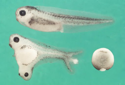
Spemann reasoned that some cells in the early embryo were able to direct the fate of other cells. By transplanting parts of one embryo to specific locations in another embryo he determined which cells acted as organizing centers, presumably by secreting regulatory molecules [see Monday's Molecule #126 and The Spemann–Mangold organizer experiment in 1924].

Here's an excerpt from the Presentation Speech.
THEME:
Nobel Laureates
Much thought has been given to the nature of the forces and causality regulating this development. It is at this point that Spemann's researches begin. He used eggs of various animal species which differ in colour, and with his simple instruments transplanted small pieces of tissue in different stages of development. By this means he was able to establish that, for example, a cell mass normally destined to become ventral epidermis - Spemann calls it presumptive ventral epidermis -could develop into nerve tissue if it were put in the place where the spinal cord was to develop. Hence, the course of development of these cells was not laid down in advance or it could - if such was the case - be altered by transplantation; so that the transplanted portion adjusted itself to its new environment. When Spemann then transplanted the anterior lip of the blastopore of an embryo into the ventral side of another embryo it grew a new brain and spinal cord. This brain and spinal cord did not arise from the transplanted cell material, but from the presumptive ventral epidermis whose course of development was thus altered by the presence of the blastopore. From this Spemann could ascertain that the blastopore had an organizing influence on its environment. The cell material which was grafted into the ventral epidermis and caused the development of the new spinal cord was actually of the kind that, developing normally, would have given rise to the notochord. Further experiments showed that it is the notochord primordia which organize the development of the primordial spinal cord, while, on the other hand, the mesoderm in the head causes the development of a primordial brain. Near this arise the so-called optic vesicles which are the origin of the retina of the eye. Where these approach the ectoderm of the head they organize the development of the lens of the eye. Or, to take another example: the anterior end of the primordial gut (the oesophagus) organizes the development of a primordial mouth and primordial teeth inside it. Thus, we now see how cell masses originally undifferentiated have the course of their development laid down by the influence of rudiments of organs formed earlier. Thereafter, a cell mass such as this can assume the role of organizer in relation to its environment.

In this way we begin to understand how the laws of development work. We begin to perceive why a primordial head arises at the anterior end of the embryo, why a brain always arises in the head and never anywhere else, or why the mouth always has its place below the primordial brain and never elsewhere.


[Image Credit: E. M. De Robertis and Hiroki Kuroda (2004)]

The images of the Nobel Prize medals are registered trademarks of the Nobel Foundation (© The Nobel Foundation). They are used here, with permission, for educational purposes only.

Tuesday, June 16, 2009

Nobel Laureate: Robert Koch


The Nobel Prize in Physiology or Medicine 1905

"for his investigations and discoveries in relation to tuberculosis"

Robert Koch (1843 - 1910) won the Noble Prize in 1905 for demonstrating that specific bacteria can cause common diseases. Tuberculosis was the specific disease mentioned in the citation.

At the time of the award, Koch was already a very famous scientist. Part of his reputation was based on The Most Famous Speech in Medical History but he was also widely respected for identifying the bacteria causes of other diseases.

The most important part of the Presentation Speech is the part that emphasizes the general contribution of Koch to the study of bacteriology (see below). Koch is recognized as one of the founders of the modern field of microbiology. One of his co-workers, Paul Ehrlich, won the Nobel Prize three years after Koch [Nobel Laureate: Paul Ehrlich].
THEME:
Nobel Laureates
To start with, developing a general methodology is as valuable as finding the correct technique for every special case. Koch's genius has blazed new trails in this respect and has given present-day research its form. To give a detailed description of this is beyond the scope of this account. I only want to mention that he had moreover already given a significant development to techniques in staining and microscopic investigation as well as in the field of experiment in his earliest work. Shortly after this he produced the important method, which is still generally the usual one, of spreading the material under investigation in a solid nutrient medium to allow each individual among the micro-organisms present to develop into a fixed colony, from which it is possible, in further research, to go on to obtain what is known as a pure culture.

Shortly after the publication of his investigations into diseases from wound infections Koch was appointed to the new Institution, the «Gesundheitsamt» (Department of Health), in Berlin. There he started work on some of the most important human diseases, namely, tuberculosis, diphtheria and typhus. He worked on the former one himself. The two latter investigations he left to his first two pupils and assistants, Loeffler and Gaffky. For all three diseases the specific bacteria were discovered and studied in detail.

To give an account of the work which Koch carried out, or accomplished through his pupils, and also to mention the work which derives more indirectly from Koch, would nearly be the same as describing the development of bacteriology over the last few decades. I will content myself with naming some of the most important discoveries and items of research which, in addition to those already named, are more directly linked with Koch's name. At the head of the German Cholera Commission Koch investigated the parasitic aetiology of cholera in Egypt and India, and discovered the cholera bacillus and the conditions necessary for its life. Experience thus gained found practical application in the development of measures taken to prevent and combat this devastating disease. In addition Koch made important investigations concerning plague in humans, malaria, tropical dysentery, and the Egyptian eye disease (trachoma) among others, and now finally concerning typhus recurrens in tropical Africa. He has also carried out work of exceptional importance, concerning a host of destructive tropical cattle diseases, such as rinderpest, Surra disease, Texas fever, and finally concerning coast fever in cattle and the trypanosome disease carried by the tsetse fly.

Through the perfection he gave to methods of culturing and identifying micro-organisms, he has been able to carry out his work with regard to disinfectants and methods of disinfection so important for practical hygiene, and advice concerning the early detection and combating of certain epidemic diseases such as cholera, typhus and malaria.

[Image Credits: photograph:zgapa.pl/drawing: Wolsztyn - Wollstein/statue: Wikipedia/movie poster: Journal of Medicine and Movies]

The images of the Nobel Prize medals are registered trademarks of the Nobel Foundation (© The Nobel Foundation). They are used here, with permission, for educational purposes only.

Thursday, June 11, 2009

Nobel Laureate: Jens Skou


The Nobel Prize in Chemistry 1997.

"for the first discovery of an ion-transporting enzyme, Na+, K+ -ATPase"


Jens C. Skou (1918 - ) won the Nobel Prize in Chemistry his work on the Na+,K+ ATPase (sodium potassium ATPase). He discovered that this membrane protein pumped sodium ions out of cells and pumped potassium ions into cells. The pump was driven by hydrolysis of ATP.

Skou shared the Nobel Prize with Paul Boyer and John Walker who worked out the mechanism of ATP synthase—the enzyme that makes ATP.

The press release describes Skou's work in some detail.
THEME:
Nobel Laureates
Na+, K+-ATPase, the first molecular pump to be discovered

It was known as early as the 1920s that the ion composition within living cells is different from that in the surroundings. Within the cells the sodium concentration is lower and the potassium concentration higher than in the liquid outside. Through the work of the Englishmen Richard Keynes and Alan Hodgkin at the beginning of the 1950s (Hodgkin received the Nobel Prize in 1963) it was known that when a nerve is stimulated sodium ions pour into the nerve cell. The difference in concentration is restored by sodium being transported out once again. That this transport required ATP was probable since the transport could be inhibited in the living cell by inhibiting the formation of ATP.

With this as the starting point Jens C. Skou searched for an ATP-degrading enzyme in the nerve membrane that could be associated with ion transport. In 1957 he published the first article on an ATPase, which was activated by sodium and potassium ions (Na + , K + -ATPase). He was the first to describe an enzyme that can promote directed (vectored) transport of substances through a cell membrane, a fundamental property of all living cells. Numerous enzymes have since been demonstrated to have essentially similar functions.

Skou used as experimental material finely ground crab nerve membranes. The ATP-degrading enzyme found in the preparation required the presence of magnesium ions and was stimulated with increasing quantities of sodium ions up to a certain limit. Above this Skou was able to obtain further stimulation if he added small quantities of potassium ions. An indication that the enzyme was coupled to the ion pump was that maximal stimulation was obtained at the concentrations of sodium and potassium that normally occur in the nerve. In his further studies of the enzyme mechanism Skou showed that sodium ions and potassium ions bind with high affinity to different places in the enzyme. In addition he showed that the phosphate group separated from ATP also binds to ATPase. This is described as a phosphorylation of the enzyme. The enzyme is dependent on sodium ions when it is phosphorylated and on potassium ions when it is dephosphorylated. Substances known to inhibit sodium/potassium transport are certain digitalis alkaloids, e.g. oubain, and Skou showed that oubain interferes in the enzyme's activation by sodium.

The picture that slowly emerged from Skou's and others' work is that the enzyme consists of two subunits, alpha and beta. The first carries the enzyme's activity and the other presumably stabilises the structure. The enzyme molecules are located in the cell membrane, often in twos, and expose surfaces to the outside as well as the inside. Three sodium ions and ATP bind to the interior surface. A phosphate is then transferred from ATP to an amino acid in the enzyme, aspartic acid, whereupon the ADP is liberated and the enzyme changes form so that the sodium ions are transported to the outside. Here they are released and two potassium ions attach instead. When the phosphorus that is bound to the enzyme is removed the potassium ions are transported into the cell and when new ATP binds to the enzyme they are rejected.

As a result of the action of the Na + , K + -ATPase, the cell keeps a high concentration of potassium in its inside. As the cell membrane is rather permeable for potassium ions, a few of these potassium ions leak out, leaving unpermeable, negative charges on the inside of the cell. Therefore, the inside of the cell membrane becomes electrically negatively charged, as compared to the outside.

This difference in potential across the membrane is necessary for a nerve stimulation to propagate along a nerve fibre or a muscle cell. This is why a shortage of nourishment or oxygen in the brain rapidly leads to unconsciousness since the ATP formation ceases and the ion pump stops. The pump is also important for maintaining cell volume. If the pump stops, the cell swells. The difference in sodium concentration between the interior and the exterior is the driving force in the uptake of important nutrients necessary to the cell, e.g. glucose and amino acids. It can also be used for transport of other ions through the cell membrane. Thus sodium ions that enter can be exchanged for calcium ions that exit.

Following the discovery of Na + , K + -ATPase other ion pumps have been discovered with similar structures and functions. Examples are Ca 2+ >-ATPase in skeletal muscle, which participates in the control of muscle contraction and H + , K + -ATPase which produces hydrochloric acid in the stomach. It is the latter enzyme that is specifically inhibited in modern treatment of stomach ulcers. Corresponding enzymes are also found in lower organisms, for example in yeast where an H + -ATPase secretes hydrogen ions formed during fermentation. As a common name these enzymes are nowadays termed P-type ATPases since they are phosphorylated during the course of the reaction.

The images of the Nobel Prize medals are registered trademarks of the Nobel Foundation (© The Nobel Foundation). They are used here, with permission, for educational purposes only.

Tuesday, June 02, 2009

Monday's Molecule #124: Winners

 
This week's winner is Mike Fraser of the University of Toronto. (Yeah, Canada!) Here's what he wrote,
The molecule is the Na+/K+ ATPase ('sodium-potassium pump'). The stoichiometry is 3Na+ out for 2K+ into the cell. In the process, ATP is converted to ADP + Pi (inorganic phosphate).

The Nobelist is Jens Skou, Chemistry 1997.
The Undergraduate winner is Jason Oakley of the University of Toronto.

The fastest correct answer was from an ineligible American but the next four correct answers were from Canadians. Maybe I should extend the ineligible delay even more!!!

Europeans and the rest of the world weren't even in the top ten.


Name this molecule. Be as specific as possible. You must also identify the missing products and reactants. Be sure to get the stoichiometry correct or it doesn't count!

Identify the Nobel Laureate who discovered this molecule.

The first person to identify the molecule plus its reactants and products and identify the Nobel Laureate, wins a free lunch at the Faculty Club. Previous winners are ineligible for six weeks from the time they first won the prize. Please note the change in the length of time you are ineligible. The idea is to give more more people a chance to win.

There are seven ineligible candidates for this week's reward: Laura Gerth of the University of Notre Dame, Stefan Tarnawsky of the University of Toronto, Dima Klenchin of the University of Wisconsin, Madison, Adam Santoro of the University of Toronto., Michael Clarkson of Waltham MA (USA), Òscar Reig of Barcelona, and Maria Altshuler of the University of Toronto.

The rest of the world has pulled ahead of the Canadians. If it wasn't for the special free lunch for people who can actually collect it, there would be no Canadian winners at all!! What's happened?

I still have one extra free lunch donated by a previous winner to a deserving undergraduate so I'm going to continue to award an additional free lunch to the first undergraduate student who can accept it. Please indicate in your email message whether you are an undergraduate and whether you can make it for lunch.

THEME:

Nobel Laureates
Send your guess to Sandwalk (sandwalk (at) bioinfo.med.utoronto.ca) and I'll pick the first email message that correctly identifies the molecule and names the Nobel Laureate(s). Note that I'm not going to repeat Nobel Prizes so you might want to check the list of previous Sandwalk postings by clicking on the link in the theme box.

Correct responses will be posted tomorrow.

Comments will be blocked for 24 hours. Comments are now open.


Thursday, May 28, 2009

Nobel Laureate: Willem Einthoven


The Nobel Prize in Physiology or Medicine 1924

"for his discovery of the mechanism of the electrocardiogram"


Willem Einthoven (1860 - 1927) won the Noble Prize in 1924 for discovering a practical machine for detecting the electrical actions of the heart. He discovered the electrocardiogram and identified its characteristic features.

Einthoven's apparatus was based on the string galvanometer, which he had developed a number of years earlier. The importance of an accurate electrocardiogram in diagnosing various heart conditions was instantly recognized. But first, the actions of a normal heart had to be carefully recorded and explained. The explanation put forth by Einthoven proved to be substantially correct.

Here's how the standard electrocardiogram is described in the Presentation Speech.
THEME:
Nobel Laureates
However, in his work in 1908 Einthoven gave an interpretation of the electrocardiogram. He starts from the fact that the stimulus (of the contraction process, the «negativity») is propagated as a wave in the muscular system of the heart. The string of the galvanometer, connected with the heart in a closed circuit in one of the usual ways, remains in the original position not only when the heart is at rest, but also when the «negativity» of the assemblage of points of the heart wall show the same value. A deflection is therefore in the first place to be expected at the beginning and at the end of a systole, and it presupposes that the condition of activity does not occur, respectively cease, simultaneously in all elements of the muscle. Further: if the contraction process (the stimulus) is propagated symmetrically in relation to the points connected to the galvanometer, then no deflection would take place either. Under such circumstances the electrocardiogram must be determined partly by the starting-point of the stimulus to the heart beat, partly by the conduction system within the heart. The point of departure for the normal heart beat has been sufficiently well known since the middle of the 1890's, the bundle of His also since that time, and Tawara's description of the ramification of the conduction system inside the ventricles known since 1906. According to Einthoven the P-peak is an expression of the propagation of the stimulus wave in the muscular system of the auricle. The negativity wave, corresponding to the stimulus wave in the His-Tawara system, is considered too weak by Einthoven to cause any deflection in the galvanometer. The QRS-complex is determined by the propagation of the stimulus wave in the muscular system of the two ventricles, proceeding in unsymmetrical fashion to the points of lead, starting at different moments at the transition of the tree-like ramified Purkinje's fibres into the various parts of the proper muscular system of the heart. When the contraction process has reached its maximum in all the points of the ventricular wall, the string returns to its original position. When the contraction ceases in the various parts at different moments, a T-peak is obtained.


It is unnecessary in this connection to consider the interpretations proposed by other investigators, as Einthoven's concept is the only one which has proved to be tenable. The interpretation that the P-peak belongs to the auricular systole is mainly based on his observation of electrocardiograms in cases of heart block in patients or during vagus stimulation in dogs. With regard to the interpretation of the QRS-complex Einthoven was evidently the first who has clearly recognized the significance of the conduction system in this connection. The train of thought in the interpretation of the T-peak can already be detected in Burdon-Sanderson's previously mentioned work.


The images of the Nobel Prize medals are registered trademarks of the Nobel Foundation (© The Nobel Foundation). They are used here, with permission, for educational purposes only.

Wednesday, May 20, 2009

Nobel Laureate: Charles Robert Richet


The Nobel Prize in Physiology or Medicine 1913

"in recognition of his work on anaphylaxis"


Charles Robert Richet (1850 - 1935) won the Noble Prize in 1913 for discovering the phenomenon known as anaphylaxis. This is a condition where the administration of an antigen causes severe symptoms, even death. Richet found that anaphylactic shock occurs only after an animal had been previously immunized and even then only after some days had passed.

It appeared as though the first immunization took several days to develop but when the process was complete a second attempt at boosting immunization causes a severe reaction. Anaphylactic shock was rare, it only happens in a small percentage of cases. We are familiar with the risk when people are known to be allergic to peanuts or insect stings.

Today we know what causes the symptoms of anaphylaxis; it's due to massive release of histamines, prostaglandins, and leukotrienes from mast cells. The release of these chemicals produces rapid heartbeat, sweating, and constriction of the airways. The symptoms can be relieved, and death prevented, by rapid treatment with epinephrine.

The primary cause of most anaphyaxis is overproduction of antigen-specific immunoglobulin E (IgE) molecules on the mast cells.1 It's the IgE molecules that interact with the antigen to cause release of histamines etc. It's not known why some antigens lead to overproduction of IgE such that subsequent exposure to the same antigen cause a massive allergic reaction. (Normal antibodies are immunoglobulin G or IgG.2)

Immunology is complicated. That's why we can't cure asthma and other allergic reactions even though the phenomena have been intensely studied for more than 100 years.

Here's an excerpt from the 1913 Presentation Speech.
THEME:
Nobel Laureates
In an age in which the leading members of the medical profession tend to concentrate on innumerable experiments demonstrating the growing immunity of the organism towards poisons already resisted successfully once, you, Sir, have found that in certain cases a completely opposite result is produced. You did not restrict yourself to this isolated observation: studied in depth by you, it has become the foundation on which you have based the evidence of a reaction that is sometimes just as regular as the phenomenon of immunity. We are not concerned solely with specific prophylaxis; thanks to you, we are now aware of a specific anaphylaxis.

We do not discount the work of those who, following your lead, have observed similar phenomena, but to you goes the honour of having established the basis of a new biological reaction, anaphylaxis, and of having been the first to demonstrate it clearly. Thereby you have opened up to medical science an enormous field of study as yet unexplored. The Staff of Professors of the Caroline Institute wishes to reward you for this achievement by conferring on you the prize instituted by our compatriot Alfred Nobel for those «who have made the most important discovery in the field of physiology or medicine».

Please accept the warm congratulations of the Institute and myself, together with the wish of us all that success will continue to crown your devoted work.


1. I do not mean to imply that IgE molecules are produced by mast cells. They are not.

2. There are several different classes (isotypes) of antibodies; IgG, IgD, IgM, IgA, and IgE. The most abundance class is IgG—that's the one most often depicted in the textbooks. It's probably the type most people think about when they think about antibodies. I did not mean to imply that the other classes are not "normal."

[Photo Credit: Wikipedia]

The images of the Nobel Prize medals are registered trademarks of the Nobel Foundation (© The Nobel Foundation). They are used here, with permission, for educational purposes only.