More Recent Comments

Showing posts with label Nobel Prize. Show all posts

Wednesday, May 13, 2009

Nobel Laureate: Richard Ernst


The Nobel Prize in Chemistry 1991.

"for his contributions to the development of the methodology of high resolution nuclear magnetic resonance (NMR) spectroscopy"


Richard R. Ernst (1933 - ) won the Nobel Prize in Chemistry for important contributions to the technology of nuclear magnetic resonance (NMR) as a tool to understanding the three-dimensional structure of molecules.

The press release describes his work in some detail.
THEME:
Nobel Laureates
Revolutionary developments make a spectroscopic technique indispensable for chemistry

The 1991 Nobel Prize in Chemistry has been awarded to Professor Richard R. Ernst of the ETH, Zurich, for important methodological developments within nuclear magnetic resonance (NMR) spectroscopy. NMR spectroscopy has during the last twenty years developed into perhaps the most important instrumental measuring technique within chemistry. This has occurred because of a dramatic increase in both the sensitivity and the resolution of the instruments, two areas in which Ernst has contributed more than anybody else.

NMR spectroscopy is today used within practically all branches of chemistry, at universities as well as industrial laboratories. The method has its most important applications as a tool for the determination of molecular structure in solution. It can today be applied to a wide variety of chemical systems, from small molecules (e.g. drugs) to proteins and nucleic acids. Further, chemists use NMR to study interactions between different molecules (e.g. enzyme - substrate, soap - water), to investigate molecular motion, to get information on the rate of chemical reactions and for many other problems. The NMR technique is today also important in related sciences, such as physics, biology and medicine.

Background

The first successful NMR experiments were reported in 1945, by two independent groups in the USA (Bloch and co-workers at Stanford and Purcell with his group at Harvard). Their discovery was awarded a Nobel Prize in Physics in 1952. The NMR phenomenon can be explained in the following way. When matter is placed in a magnetic field, some of the atomic nuclei (e.g. nuclei of hydrogen atoms, called protons) behave like microscopic compass needles. These tiny compass needles (called nuclear spins) can, according to the laws of quantum mechanics, orient themselves with respect to the magnetic field in only a few ways. These orientations are characterized by different energy levels. The nuclear spins can be forced to jump between levels if the sample is exposed to radio waves of exactly specified frequency. The frequency is varied during the course of the experiment and, when it exactly matches the characteristic frequency of the nuclei (the resonance frequency), an electric signal is induced in the detector. The strength of the signal is plotted as a function of frequency in a diagram called the NMR spectrum. Around 1950, it was discovered that nuclear resonance frequencies depended not only on the nature of the atomic nuclei, but also on their chemical environment. The possibility of using NMR as a tool for chemical analysis soon became obvious and was mentioned by, among others, Professor Purcell in his 1952 Nobel lecture. A fundamental difficulty in the early days was the relatively low sensitivity of the NMR method.

A major breakthrough occurred in 1966 when Ernst (together with Weston A. Anderson, USA) discovered that the sensitivity of NMR spectra could be increased dramatically if the slow radiofrequency sweep that the sample was exposed to was replaced by short and intense radiofrequency pulses. The signal was then measured as a function of time after the pulse. The next pulse and signal acquisition were started after a few seconds, and the signals after each pulse were summed in a computer. The NMR signal measured as a function of time is not amenable to a simple interpretation (see Figure la). It is however possible to analyze what resonance frequencies are present in such a signal - and to convert it to an NMR spectrum - by a mathematical operation (Fourier transformation, FT) performed rapidly in the computer. The result of the Fourier transformation of Figure la is shown in Figure lb.

This discovery is the basis of modern NMR spectroscopy. The ten-fold, and sometimes hundred-fold, increase in sensitivity has made it possible to study small amounts of material as well as chemically interesting isotopes of low natural occurrence, e.g. carbon- 13. The enormous potential of the new technique - called FT NMR - quickly became obvious to NMR spectroscopists. The chemical research community got access to it in the early seventies through commercial FT NMR instruments. Nowadays, practically no other types of NMR spectrometer are manufactured.

By the end of the sixties, NMR spectroscopists had begun to use new magnet designs, based on superconducting materials, and the quality of spectra - expressed both in terms of sensitivity and resolution - improved quickly during the seventies. Consequently, more complex systems could be studied and more sophishcated questions answered. To move to very large molecules, macromolecules, another breakthrough was necessary, and this again carried the signature of Ernst. Inspired by a lecture of Jean Jeener, Belgium, at a summer school at the beginning of the seventies, Ernst and co-workers showed in 1975-76 how "two-dimensional" (2D) NMR experiments could be performed and demonstrated that 2D FT NMR opened entirely new possibilities for chemical research.

This 2D methods functions in the following way. Nuclear spins in a magnetic field are now subjected to sequences of radio-frequency pulses rather than to single pulses. The time course of the experiment is divided into four intervals. During the "preparation period", the equilibrium of the nuclear spin system is distorted by one or several pulses. This non-equilibrium is allowed to evolve for a certain time (the "evolution period"), after which the next series of pulses (the "mixing period") leads to the "detection period". Here the NMR signal is detected as a function of time in the same way as in ordinary, one-dimensional FT NMR. After this, one moves to the next preparation period and repeats the experiment with different evolution period. The change in the evolution period causes the signal measured during the detection period to change. One might say that the history of spins during the evolution period becomes encoded in the variation of the signal measured during the detection period. This gives a two-dimensional table with signal intensity as a function of both the point in time during the detection period and the length of the evolution period. Finally, the Fourier transformation is performed twice - with respect to both these time parameters - to obtain a two-dimensional frequency spectrum in the form of a map of the dependence of the signal intensity on two frequency parameters (denoted f1 and f2 in Figure 2).

Introduction of the second frequency dimension allows the spectral information to attain much higher resolution - like looking at the skyline of a mountain range and then looking at the whole range from an aircraft above. Depending on the design of the preparation and the mixing periods, one obtains a variety of 2D NMR experiments. Some are used to spread the information over two dimensions rather than one (separation of interactions) while others are designed to find which nuclei have some form of contact with each other (correlation of signals).

In the mid-seventies, Ernst also proposed a method of obtaining NMR-tomographic images which became one of the most common (the NMR tomography method as such was earlier realized by Lauterbur in the USA, Mansfield in England and others).

Since the mid-seventies, Ernst and co-workers have continuously and decisively contributed to the development of NMR spectroscopy, and in particular its two-, and more recently three- and multi-dimensional varieties. Applications of his methods were soon to come. For example, it has become possible over the past ten years to use NMR to determine the three-dimensional structure of organic and inorganic compounds as well as proteins and other biological macromolecules in solution with an accuracy comparable to what can be attained in crystals using X-ray diffraction. Interactions between biological molecules and other substances (metal ions, water, drugs) have also been studied in detail. Other important chemical applications are identification of chemical species (where NMR spectra act as the fingerprint of a molecule), studies of rates of certain chemical reactions and of molecular motions in the liquid state. In the border area between chemistry and biology, NMR is being used to study how metabolic processes are influenced by drugs, ischaemia etc. Ernst's own work often falls in the border area between chemistry and physics and can, if one so wishes, be treated as extremely elegant experimental verification of the correctness of quantum mechanics.

[Photo Credit: Science Festival]

The images of the Nobel Prize medals are registered trademarks of the Nobel Foundation (© The Nobel Foundation). They are used here, with permission, for educational purposes only.

Nobel Laureate: Harald zur Hausen


The Nobel Prize in Physiology or Medicine 2008

"for his discovery of human papilloma viruses causing cervical cancer"


Harald zur Hausen (1936 - ) won the Noble Prize in 2008 for discovering that a virus, human papilloma virus, causes cervical cancer. He also won a Gairdner Award in 2008.

Zur Hausen's discovery led eventually to the development of an HPV vaccine. Gardasil is the best known of the two vaccines on the market. Most doctors recommend that young girls be vaccinated.

Here's the 2008 press release on Zur Hausen.
THEME:
Nobel Laureates
Discovery of human papilloma virus causing cervical cancer

Against the prevailing view during the 1970s, Harald zur Hausen postulated a role for human papilloma virus (HPV) in cervical cancer. He assumed that the tumour cells, if they contained an oncogenic virus, should harbour viral DNA integrated into their genomes. The HPV genes promoting cell proliferation should therefore be detectable by specifically searching tumour cells for such viral DNA. Harald zur Hausen pursued this idea for over 10 years by searching for different HPV types, a search made difficult by the fact that only parts of the viral DNA were integrated into the host genome. He found novel HPV-DNA in cervix cancer biopsies, and thus discovered the new, tumourigenic HPV16 type in 1983. In 1984, he cloned HPV16 and 18 from patients with cervical cancer. The HPV types 16 and 18 were consistently found in about 70% of cervical cancer biopsies throughout the world.

Importance of the HPV discovery

The global public health burden attributable to human papilloma viruses is considerable. More than 5% of all cancers worldwide are caused by persistent infection with this virus. Infection by the human papilloma virus is the most common sexually transmitted agent, afflicting 50-80% of the population. Of the more than 100 HPV types known, about 40 infect the genital tract, and 15 of these put women at high risk for cervical cancer. In addition, HPV is found in some vulval, penile, oral and other cancers. Human papilloma virus can be detected in 99.7% of women with histologically confirmed cervical cancer, affecting some 500,000 women per year.

Harald zur Hausen demonstrated novel properties of HPV that have led to an understanding of mechanisms for papilloma virus-induced carcinogenesis and the predisposing factors for viral persistence and cellular transformation. He made HPV16 and 18 available to the scientific community. Vaccines were ultimately developed that provide ≥95 % protection from infection by the high risk HPV16 and 18 types. The vaccines may also reduce the need for surgery and the global burden of cervical cancer.


[Photo Credit: IBMLive]

The images of the Nobel Prize medals are registered trademarks of the Nobel Foundation (© The Nobel Foundation). They are used here, with permission, for educational purposes only.

Friday, May 01, 2009

Nobel Laureate: Paul Ehrlich


The Nobel Prize in Physiology or Medicine 1908

"in recognition of their work on immunity"


Paul Ehrlich (1854 - 1915) won the Noble Prize in 1908 for his contributions to understanding immunology. His co-recipient was Ilya Ilyich Mechnikov.

Ehrlich was already a well-known scientist at the time he received that Nobel Prize and he subsequently went on to achieve even greater fame for synthesizing a drug to treat syphilis [Monday's Molecule #119].

Although Ehrlich's specific contributions to immunology aren't mentioned in the presentation speech, they mostly concern the discovery of antibodies. Here's how his contribution is described ...
THEME:
Nobel Laureates
An endless series of questions now arises: Why are antibodies only built up against some substances and not against all substances which are foreign to the organism? Where are the antibodies formed? By what process are they formed? What is the nature and constitution of these antibodies? How do they react on the microorganisms and their poisons? And various other questions which are important as regards the development and practical utilization of the theory of immunity. It is also a matter of great interest that connecting links have been found between the theory of immunity and the normal physiological processes.

A great deal of intensive and very fruitful work has been devoted to these questions in the last one and a half decades. A large number of research scientists have served the cause of science well by their discoveries and achievements. It is not possible here to report on the extent to which the questions have been answered, neither is it possible to describe the separate accomplishments of individual scientists in this field.

A man who has been responsible for important scientific progress as organizer and leader in this field deserves to be mentioned among the first of those who have dedicated themselves to a study of immunity, is the research scientist Paul Ehrlich, already famous for his other biological work, and the Professorial Staff of the Caroline Institute wishes to honour him too with this year's Nobel Prize for his work in the sphere of immunity.


[Photo Credit: Wellcome Trust Photographic Library]

The images of the Nobel Prize medals are registered trademarks of the Nobel Foundation (© The Nobel Foundation). They are used here, with permission, for educational purposes only.

Thursday, April 23, 2009

Nobel Laureate: Sir Paul Nurse


The Nobel Prize in Physiology or Medicine 2001

"for their discoveries of key regulators of the cell cycle"

Sir Paul M. Nurse (1949 - ) won the Noble Prize in 2001 for his contributions to understanding the regulation of gene expression in yeast cells. His co-recipients were Leland Hartwell and Tim Hunt.

A major part of most signaling pathways is the phosphorylation of proteins. The attachment of a phosphate group to a protein can convert it from an active state to an inactive state, or vice versa. Enzymes that catalyze phosphorylations are called "kinases" and one of the most common kinases is cyclin-dependent kinase or CDK. The activity of the kinase is itself regulated by proteins called cyclins.

Cyclins are produced at various stages of the cell cycle as it progresses from a growth state through mitosis and cell division as shown below in the press release. Paul Nurse's contribution to understanding the cell cycle was to characterize the cyclin-dependent kinase.

THEME:
Nobel Laureates
Press Release

Summary

All organisms consist of cells that multiply through cell division. An adult human being has approximately 100 000 billion cells, all originating from a single cell, the fertilized egg cell. In adults there is also an enormous number of continuously dividing cells replacing those dying. Before a cell can divide it has to grow in size, duplicate its chromosomes and separate the chromosomes for exact distribution between the two daughter cells. These different processes are coordinated in the cell cycle.

This year's Nobel Laureates in Physiology or Medicine have made seminal discoveries concerning the control of the cell cycle. They have identified key molecules that regulate the cell cycle in all eukaryotic organisms, including yeasts, plants, animals and human. These fundamental discoveries have a great impact on all aspects of cell growth. Defects in cell cycle control may lead to the type of chromosome alterations seen in cancer cells. This may in the long term open new possibilities for cancer treatment.

Leland Hartwell (born 1939), Fred Hutchinson Cancer Research Center, Seattle, USA, is awarded for his discoveries of a specific class of genes that control the cell cycle. One of these genes called "start" was found to have a central role in controlling the first step of each cell cycle. Hartwell also introduced the concept "checkpoint", a valuable aid to understanding the cell cycle.

Paul Nurse (born 1949), Imperial Cancer Research Fund, London, identified, cloned and characterized with genetic and molecular methods, one of the key regulators of the cell cycle, CDK (cyclin dependent kinase). He showed that the function of CDK was highly conserved during evolution. CDK drives the cell through the cell cycle by chemical modification (phosphorylation) of other proteins.

Timothy Hunt (born 1943), Imperial Cancer Research Fund, London, is awarded for his discovery of cyclins, proteins that regulate the CDK function. He showed that cyclins are degraded periodically at each cell division, a mechanism proved to be of general importance for cell cycle control.

One billion cells per gram tissue

Cells having their chromosomes located in a nucleus and separated from the rest of the cell, so called eukaryotic cells, appeared on earth about two billion years ago. Organisms consisting of such cells can either be unicellular, such as yeasts and amoebas, or multi-cellular such as plants and animals. The human body consists of a huge number of cells, on the average about one billion cells per gram tissue. Each cell nucleus contains our entire hereditary material (DNA), located in 46 chromosomes (23 pairs of chromosomes).

It has been known for over one hundred years that cells multiply through division. It is however only during the last two decades that it has become possible to identify the molecular mechanisms that regulate the cell cycle and thereby cell division. These fundamental mechanisms are highly conserved through evolution and operate in the same manner in all eukaryotic organisms.

The phases of the cell cycle

The cell cycle consists of several phases (see figure). In the first phase (G1) the cell grows and becomes larger. When it has reached a certain size it enters the next phase (S), in which DNA-synthesis takes place. The cell duplicates its hereditary material (DNA-replication) and a copy of each chromosome is formed. During the next phase (G2) the cell checks that DNA-replication is completed and prepares for cell division. The chromosomes are separated (mitosis, M) and the cell divides into two daughter cells. Through this mechanism the daughter cells receive identical chromosome set ups. After division, the cells are back in G1 and the cell cycle is completed.

The duration of the cell cycle varies between different cell types. In most mammalian cells it lasts between 10 and 30 hours. Cells in the first cell cycle phase (G1) do not always continue through the cycle. Instead they can exit from the cell cycle and enter a resting stage (G0).

Cell cycle control

For all living eukaryotic organisms it is essential that the different phases of the cell cycle are precisely coordinated. The phases must follow in correct order, and one phase must be completed before the next phase can begin. Errors in this coordination may lead to chromosomal alterations. Chromosomes or parts of chromosomes may be lost, rearranged or distributed unequally between the two daughter cells. This type of chromosome alteration is often seen in cancer cells.

It is of central importance in the fields of biology and medicine to understand how the cell cycle is controlled. This year's Nobel Laureates have made seminal discoveries at the molecular level of how the cell is driven from one phase to the next in the cell cycle.

Cell cycle genes in yeast cells

Leland Hartwell realized already at the end of the 1960s the possibility of studying the cell cycle with genetic methods. He used baker's yeast, Saccharomyces cerevisiae, as a model system, which proved to be highly suitable for cell cycle studies. In an elegant series of experiments 1970-71, he isolated yeast cells in which genes controlling the cell cycle were altered (mutated). By this approach he succeeded to identify more than one hundred genes specifically involved in cell cycle control, so called CDC-genes (cell division cycle genes). One of these genes, designated CDC28 by Hartwell, controls the first step in the progression through the G1-phase of the cell cycle, and was therefore also called "start".

In addition, Hartwell studied the sensitivity of yeast cells to irradiation. On the basis of his findings he introduced the concept checkpoint, which means that the cell cycle is arrested when DNA is damaged. The purpose of this is to allow time for DNA repair before the cell continues to the next phase of the cycle. Later Hartwell extended the checkpoint concept to include also controls ensuring a correct order between the cell cycle phases.

A general principle

Paul Nurse followed Hartwell's approach in using genetic methods for cell cycle studies. He used a different type of yeast, Schizosaccharomyces pombe, as a model organism. This yeast is only distantly related to baker's yeast, since they separated from each other during evolution more than one billion years ago.

In the middle of the 1970s, Paul Nurse discovered the gene cdc2 in S. pombe. He showed that this gene had a key function in the control of cell division (transition from G2 to mitosis, M). Later he found that cdc2 had a more general function. It was identical to the gene ("start") that Hartwell earlier had identified in baker's yeast, controlling the transition from G1 to S.

This gene (cdc2) was thus found to regulate different phases of the cell cycle. In 1987 Paul Nurse isolated the corresponding gene in humans, and it was later given the name CDK1 (cyclin dependent kinase 1). The gene encodes a protein that is a member of a family called cyclin dependent kinases, CDK. Nurse showed that activation of CDK is dependent on reversible phosphorylation, i.e. that phosphate groups are linked to or removed from proteins. On the basis of these findings, half a dozen different CDK molecules have been found in humans.

The discovery of the first cyclin

Tim Hunt discovered the first cyclin molecule in the early 1980s. Cyclins are proteins formed and degraded during each cell cycle. They were named cyclins because the levels of these proteins vary periodically during the cell cycle. The cyclins bind to the CDK molecules, thereby regulating the CDK activity and selecting the proteins to be phosphorylated.

The discovery of cyclin, which was made using sea urchins, Arbacia, as a model system, was the result of Hunt's finding that this protein was degraded periodically in the cell cycle. Periodic protein degradation is an important general control mechanism of the cell cycle. Tim Hunt later discovered cyclins in other species and found that also the cyclins were conserved during evolution. Today around ten different cyclins have been found in humans.

The engine and the gear box of the cell cycle

The three Nobel Laureates have discovered molecular mechanisms that regulate the cell cycle. The amount of CDK-molecules is constant during the cell cycle, but their activities vary because of the regulatory function of the cyclins. CDK and cyclin together drive the cell from one cell cycle phase to the next. The CDK-molecules can be compared with an engine and the cyclins with a gear box controlling whether the engine will run in the idling state or drive the cell forward in the cell cycle.

A great impact of the discoveries

Most biomedical research areas will benefit from these basic discoveries, which may result in broad applications within many different fields. The discoveries are important in understanding how chromosomal instability develops in cancer cells, i.e. how parts of chromosomes are rearranged, lost or distributed unequally between daughter cells. It is likely that such chromosome alterations are the result of defective cell cycle control. It has been shown that genes for CDK-molecules and cyclins can function as oncogenes. CDK-molecules and cyclins also collaborate with the products of tumour suppressor genes (e.g. p53 and Rb) during the cell cycle.

The findings in the cell cycle field are about to be applied to tumour diagnostics. Increased levels of CDK-molecules and cyclins are sometimes found in human tumours, such as breast cancer and brain tumours. The discoveries may in the long term also open new principles for cancer therapy. Already now clinical trials are in progress using inhibitors of CDK-molecules.


The different phases of the cell cycle. In the first phase (G1) the cell grows. When it has reached a certain size it enters the phase of DNA-synthesis (S) where the chromosomes are duplicated. During the next phase (G2) the cell prepares itself for division. During mitosis (M) the chromosomes are separated and segregated to the daughter cells, which thereby get exactly the same chromosome set up. The cells are then back in G1 and the cell cycle is completed.

This year's Nobel Laureates, using genetic and molecular biology methods, have discovered mechanisms controlling the cell cycle. CDK-molecules and cyclins drive the cell from one phase to the next. The CDK-molecules can be compared with an engine and the cyclins with a gear box controlling whether the engine will run in the idling state or drive the cell forward in the cell cycle.


[Photo Credit: Havard University]

The images of the Nobel Prize medals are registered trademarks of the Nobel Foundation (© The Nobel Foundation). They are used here, with permission, for educational purposes only.

Thursday, April 16, 2009

Nobel Laureate: Max Theiler


The Nobel Prize in Physiology or Medicine 1951

"for his discoveries concerning yellow fever and how to combat it"


Max Theiler (1899 - 1972) won the Noble Prize in 1951 for his work on combating yellow fever.

Theiler's most important contribution was the discovery of a variant of the yellow fever virus that did not cause the disease in humans. When injected into healthy patients, this variant produced immunity to the normal disease-producing virus.

This discovery was not immediately useful since attenuated virus from mice was more effective in producing immunity—a result also discovered by Theiler. The Nobel Committee felt that Theiler had made a significant contribution to understanding viral diseases.

One gets the impression from reading the presentation speech that Theiler was also being recognized as a representative of work done by the Rockefeller Foundation.

THEME:
Nobel Laureates
The significance of Max Theiler's discovery must be considered to be very great from the practical point of view, as effective protection against yellow fever is one condition for the development of the tropical regions - an important problem in an overpopulated world. Dr. Theiler's discovery does not imply anything fundamentally new, for the idea of inoculation against a disease by the use of a variant of the etiologic agent which, though harmless, produces immunity, is more than 150 years old. Jenner used a natural virus variant, cowpox virus, against smallpox, and Pasteur produced a similar variant of the rabies virus by repeated passage through animals. So far there have been only a few successful attempts to master a disease by such measures, but Dr. Theiler's discovery gives new hope that in this manner we shall succeed in mastering other virus diseases, many of which have a devastating, effect and against which we are still entirely powerless. Max Theiler, therefore, has rendered mankind such a service as Nobel made a condition for the awarding of this prize.

Dr. Theiler. For a period of almost forty years the International Health Division of the Rockefeller Foundation has carried on very comprehensive and fruitful work in combating yellow fever and extending our knowledge of it. Among the many who have made their contributions, you take an especially prominent place, because you have made their contributions profitable and because you have opened the way to greater understanding of the epidemiology of the disease and to an effective prophylaxis against it. The Caroline Institute esteems your research work so highly, not the least for its practical value, that it has found it proper to award this year's Nobel Prize in Physiology or Medicine to you.

I ask you, Dr. Theiler, to receive the prize from the hands of His Majesty, our gracious King.


[Photo Credit: ©Bettmann/CORBIS, Rights Managed, Corbis]

The images of the Nobel Prize medals are registered trademarks of the Nobel Foundation (© The Nobel Foundation). They are used here, with permission, for educational purposes only.

Wednesday, April 08, 2009

Nobel Laureates: Mario Capecchi, Martin Evans, and Oliver Smithies


The Nobel Prize in Physiology or Medicine 2007

"for their discoveries of principles for introducing specific gene modifications in mice by the use of embryonic stem cells"


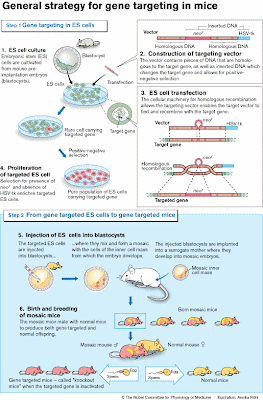
Mario R. Capecchi (1937 - ), Sir Martin J. Evans (1941 - ), and Oliver Smithies (1925 - ) won the Noble Prize in 2007 for developing techniques to transform embryonic stem cells with foreign genes integrated at a specific place in the genome, then using those cells to make transgenic mice.

The Press Release describing this work is a well-written description of how the techniques was developed.

This is how to make knock-out mice.
THEME:
Nobel Laureates
Summary

This year's Nobel Laureates have made a series of ground-breaking discoveries concerning embryonic stem cells and DNA recombination in mammals. Their discoveries led to the creation of an immensely powerful technology referred to as gene targeting in mice. It is now being applied to virtually all areas of biomedicine – from basic research to the development of new therapies.

Gene targeting is often used to inactivate single genes. Such gene "knockout" experiments have elucidated the roles of numerous genes in embryonic development, adult physiology, aging and disease. To date, more than ten thousand mouse genes (approximately half of the genes in the mammalian genome) have been knocked out. Ongoing international efforts will make "knockout mice" for all genes available within the near future.

With gene targeting it is now possible to produce almost any type of DNA modification in the mouse genome, allowing scientists to establish the roles of individual genes in health and disease. Gene targeting has already produced more than five hundred different mouse models of human disorders, including cardiovascular and neuro-degenerative diseases, diabetes and cancer.

Modification of genes by homologous recombination

Information about the development and function of our bodies throughout life is carried within the DNA. Our DNA is packaged in chromosomes, which occur in pairs – one inherited from the father and one from the mother. Exchange of DNA sequences within such chromosome pairs increases genetic variation in the population and occurs by a process called homologous recombination. This process is conserved throughout evolution and was demonstrated in bacteria more than 50 years ago by the 1958 Nobel Laureate Joshua Lederberg.

Mario Capecchi and Oliver Smithies both had the vision that homologous recombination could be used to specifically modify genes in mammalian cells and they worked consistently towards this goal.

Capecchi demonstrated that homologous recombination could take place between introduced DNA and the chromosomes in mammalian cells. He showed that defective genes could be repaired by homologous recombination with the incoming DNA. Smithies initially tried to repair mutated genes in human cells. He thought that certain inherited blood diseases could be treated by correcting the disease-causing mutations in bone marrow stem cells. In these attempts Smithies discovered that endogenous genes could be targeted irrespective of their activity. This suggested that all genes may be accessible to modification by homologous recombination.

Embryonic stem cells – vehicles to the mouse germ line

The cell types initially studied by Capecchi and Smithies could not be used to create gene-targeted animals. This required another type of cell, one which could give rise to germ cells. Only then could the DNA modifications be inherited.

Martin Evans had worked with mouse embryonal carcinoma (EC) cells, which although they came from tumors could give rise to almost any cell type. He had the vision to use EC cells as vehicles to introduce genetic material into the mouse germ line. His attempts were initially unsuccessful because EC cells carried abnormal chromosomes and could not therefore contribute to germ cell formation. Looking for alternatives Evans discovered that chromosomally normal cell cultures could be established directly from early mouse embryos. These cells are now referred to as embryonic stem (ES) cells.

The next step was to show that ES cells could contribute to the germ line (see Figure). Embryos from one mouse strain were injected with ES cells from another mouse strain. These mosaic embryos (i.e. composed of cells from both strains) were then carried to term by surrogate mothers. The mosaic offspring was subsequently mated, and the presence of ES cell-derived genes detected in the pups. These genes would now be inherited according to Mendel’s laws.

Evans now began to modify the ES cells genetically and for this purpose chose retroviruses, which integrate their genes into the chromosomes. He demonstrated transfer of such retroviral DNA from ES cells, through mosaic mice, into the mouse germ line. Evans had used the ES cells to generate mice that carried new genetic material.

Two ideas come together – homologous recombination in ES cells

By 1986 all the pieces were at hand to begin generating the first gene targeted ES cells. Capecchi and Smithies had demonstrated that genes could be targeted by homologous recombination in cultured cells, and Evans had contributed the necessary vehicle to the mouse germ line – the ES-cells. The next step was to combine the two.

For their initial experiments both Smithies and Capecchi chose a gene (hprt) that was easily identified. This gene is involved in a rare inherited human disease (Lesch-Nyhan syndrome). Capecchi refined the strategies for targeting genes and developed a new method (positive-negative selection, see Figure) that could be generally applied.

Birth of the knockout mouse – the beginning of a new era in genetics

The first reports in which homologous recombination in ES cells was used to generate gene-targeted mice were published in 1989. Since then, the number of reported knockout mouse strains has risen exponentially. Gene targeting has developed into a highly versatile technology. It is now possible to introduce mutations that can be activated at specific time points, or in specific cells or organs, both during development and in the adult animal.

Gene targeting is used to study health and disease

Almost every aspect of mammalian physiology can be studied by gene targeting. We have consequently witnessed an explosion of research activities applying the technology. Gene targeting has now been used by so many research groups and in so many contexts that it is impossible to make a brief summary of the results. Some of the later contributions of this year's Nobel Laureates are presented below.

Gene targeting has helped us understand the roles of many hundreds of genes in mammalian fetal development. Capecchis research has uncovered the roles of genes involved in mammalian organ development and in the establishment of the body plan. His work has shed light on the causes of several human inborn malformations.

Evans applied gene targeting to develop mouse models for human diseases. He developed several models for the inherited human disease cystic fibrosis and has used these models to study disease mechanisms and to test the effects of gene therapy.

Smithies also used gene targeting to develop mouse models for inherited diseases such as cystic fibrosis and the blood disease thalassemia. He has also developed numerous mouse models for common human diseases such as hypertension and atherosclerosis.

In summary, gene targeting in mice has pervaded all fields of biomedicine. Its impact on the understanding of gene function and its benefits to mankind will continue to increase over many years to come.

[Photo Credits: Mario Capecchi: Reuters,DayLife, Sir Martin J. Evans: Reuters, DayLife, Oliver Smithies: University of North Carolina, Chapel Hill.]

The images of the Nobel Prize medals are registered trademarks of the Nobel Foundation (© The Nobel Foundation). They are used here, with permission, for educational purposes only.

Wednesday, April 01, 2009

Nobel Laureate: Adolf von Baeyer


The Nobel Prize in Chemistry 1905.

"in recognition of his services in the advancement of organic chemistry and the chemical industry, through his work on organic dyes and hydroaromatic compounds"


Johann Friedrich Wilhelm Adolf von Baeyer (1835 - 1917) won the Nobel Prize in Chemistry for his work on the preparation of organic dyes from coal tar.

His most notable achievement was the synthesis of indigo dye and determination of its structure. A cheap industrial synthesis of indigo was soon developed, freeing Europe from its dependence on indigo from India.

He was also the first person to synthesize phenolphthalein, the well-known acid or base indicator.

The presentation speech highlights the importance of the relationship between basic science and industry.
THEME:
Nobel Laureates
The complex and unique composition of indigo, however, made this also one of the hardest of tasks. Here there could be no question of one of those casual discoveries, which by happy accident seem to achieve half the work. Years of work were required for even von Baeyer's acumen and experimental skill to achieve the necessary insight into the pigment's chemical composition and to be able to manufacture it from simpler constituents. Even after the purely scientific part of the work had been completed it still took a number of years to make the results obtained from research applicable to technology.

Von Baeyer succeeded in producing indigo synthetically in three principal ways, namely from ortho-nitrophenylacetic acid, from ortho-nitrocinnamic acid and from ortho-nitrobenzaldehyde and acetone. This paved the way for the reproduction of indigo from raw material obtainable without much difficulty from coal tar. And if the problem of producing indigo industrially has now been solved from the technical as well as the economic point of view, this is entirely due to von Baeyer's basic work in the fields in question.


The images of the Nobel Prize medals are registered trademarks of the Nobel Foundation (© The Nobel Foundation). They are used here, with permission, for educational purposes only.

Wednesday, March 25, 2009

Nobel Laureate: Selman Waksman


The Nobel Prize in Physiology or Medicine 1952

"for his discovery of streptomycin, the first antibiotic effective against tuberculosis"

Selman Abraham Waksman (1888 - 1973) won the Noble Prize in 1952. The award was for discovering streptomycin.

Waksman was a soil microbiologist at Rutgers University in New Jersey (USA). In the 1930s, after the success of penicillin, he decided to change the focus of his research and look for more antibiotics. He reasoned that soil microorganisms should be a good source of novel anti-bacterial drugs.

Streptomycin was the most famous of the many antibiotics discovered in Waksman's lab. It was largely due to the dedicated work of a graduate student, Albert Schatz, who first identified streptomycin's potent effect on gram negative bacteria in October 1943. Over the next few years, Waksman became famous for discovering streptomycin and Schatz was all but forgotten.

In 1950, Schatz sued his former supervisor for recognition, and a share of the royalties. The case was settled out of court with Rutgers agreeing that Schatz and Waksman would be identified as co-discoverers of streptomycin. Schatz received a share of the royalties.

In spite of this settlement, the Nobel Prize committee awarded the prize to Wakesman and not to Waksman and Schatz. This was mildly controversial at the time but didn't qualify as a major scandal. It seems more egregious today.

The issue is part of a continuing controversy about how to attribute recognition when graduate students are working under the direction of their supervisors. There's no better way to start a fight than to bring this up with a group of graduate students. Are they apprentices, slaves, or collaborators?

I am indebted to Philip Johnson of York University (Toronto, Canada) for alerting me to the controversy and for sending along this excellent article about Albert Schatz.

Waksman does not specifically mention Schatz's contribution in his Nobel lecture but he is mentioned in the presentation speech (see below) in an obvious attempt to minimize his contribution. Knowing what we know now, should the Nobel Prize website be modified to include a discussion of the controversy? I think it should.

THEME:
Nobel Laureates
In 1940 Dr.Waksman and his collaborator had succeeded in isolating the first antibiotic, which was called «actinomycin» and it was very toxic. In 1942 another antibiotic was detected and studied, called «streptothricin». This had a high degree of activity against many bacteria and also against the tubercle bacillus. Further studies revealed that streptothricin was too toxic. During the streptothricin studies Dr. Waksman and his collaborators developed a series of test-methods, which turned out to be very useful in the isolation of streptomycin in 1943.

Encouraged by the discovery of streptothricin and stimulated by the triumphal development of penicillin treatment, the research team headed by Dr.Waksman continued their untiring search for new antibiotic-producing microbes. Before the discovery of streptomycin no less than 10,000 different soil microbes had been studied for their antibiotic activity. Dr. Waksman directed this work and distributed the various lines of research among his young assistants. One of these was Albert Schatz, who had previously worked with Dr. Waksman for 2 months and in June 1943 returned to the laboratory. Dr. Waksman gave him the task of isolating new species of Actinomyces. After a few months he isolated two strains of Actinomyces which were shown to be identical with Streptomyces griseus, discovered by Dr. Waksman in 1915. In contrast to the previous one the rediscovered microbe was shown to have antibiotic activity. To this antibiotic Dr. Waksman gave the name «streptomycin». He studied the antibiotic effect of streptomycin with Schatz and Bugie and found that it was active against several bacteria including the tubercle bacillus. These preliminary studies were completed in a relatively short time, thanks to the clear principles which had been set out previously by Dr. Waksman for the study of streptothricin.


The images of the Nobel Prize medals are registered trademarks of the Nobel Foundation (© The Nobel Foundation). They are used here, with permission, for educational purposes only.
Thursday, March 19, 2009

Nobel Laureate: Willard Libby


The Nobel Prize in Chemistry 1960.

"for his method to use carbon-14 for age determination in archaeology, geology, geophysics, and other branches of science"


Willard Frank Libby (1908 - 1980) won the Nobel Prize in Chemistry for using 14C decay to date organic material. Libby set out to study cosmic rays. He, and others, determined that one of the effects of cosmic ray is to produce carbon-14 atoms from nitrogen-14 atoms in the upper atmosphere.

He determined that the rate of production of carbon-14 and its rate of disintegration (half-life ~5600 years1) has reached an equilibrium. No matter where you find carbon, in the ocean, the atmosphere, or the biosphere, its radioactivity corresponds to about 14 disintegrations per minute per gram.

Living things incorporate this equilibrium mixture of 14C and 12C. Thus, we, like all other living things, are radioactive and this level of radioactivity can be measured using techniques that Willard Libby developed. When living things die, they stop incorporating carbon and the existing 14C continues to decay. As time goes on, the level of radioactivity declines with a half-life of ~5600 years. The age of organic material can be determined directly by measuring the remaining radioactivity of extracted carbon.

That's the basis of radiocarbon dating. Libby confirmed the feasibility of the technique by dating Egyptian artifacts, tree rings of known age, and the dead sea scrolls (labeled "Bible" in the figure). The results confirmed that radiocarbon dating works.


The results were published in the late 1940's. Since then, the technology has improved considerably. Today, scientists measure 14C directly using mass spectrometry so they don't have to wait for it to decay. Detailed calibration curves have been worked out to take account of the fact that cosmic ray intensity has varied somewhat over the past few thousand years.

With current technology, reliable dates back as far as 60,000 years can be obtained. This is about the limit of radiocarbon dating because the half-life of Carbon-14 is so short compared to more long-lived isotopes.

The presentation speech highlights the importance of radiocarbon dating in a number of disciplines.
THEME:
Nobel Laureates
Libby's dating method soon attracted attention from the scientific world, and it was not long before carbon-14 laboratories were set up in many countries. Today, some forty institutions carry on investigations in this field, nearly half of them in America. Also here, in Sweden, we have such institutions, and their investigations have given results of great value. All age determinations - nowadays several thousand every year - are published in a general review, and thus made rapidly available throughout the world. The literature in this field has grown from year to year, and at present covers an impressive area.

One of the scientists who suggested Libby as a candidate for the Nobel Prize has characterized his work in the following way: "Seldom has a single discovery in chemistry had such an impact on the thinking in so many fields of human endeavour. Seldom has a single discovery generated such wide public interest".

Professor Libby. The idea you had 13 years ago of trying to determine the age of biological materials by measuring their carbon-14 activity was a brilliant impulse. Thanks to your great experimental skill, acquired during many years devoted to the study of weakly radioactive substances, you have succeeded in developing a method that is indispensible for research work in many fields and in many institutes throughout the world. Archaeologists, geologists, geophysicists, and other scientists are greatly indebted to you for the valuable support you have given them in their work. The Swedish Academy of Sciences desires to join those who offer you grateful thanks for what you have done for the benefit of so many sciences, and has decided to award you this year's Nobel Prize for Chemistry. May I congratulate you on behalf of the Academy, and ask you to receive the prize from the hands of His Majesty the King.


1. The modern value is 5730±40 years.

[Photo Credit: University of California History Digital Archives, Copyright © 2006 The Regents of the University of California.]

The images of the Nobel Prize medals are registered trademarks of the Nobel Foundation (© The Nobel Foundation). They are used here, with permission, for educational purposes only.

Monday, March 16, 2009

Monday's Molecule #112

 
You may have noticed that today's molecule isn't a molecule. Your task is to identify what this equation is describing.

There's only one Nobel Laureate whose discovery is relevant.

The first person to identify the equation and the Nobel Laureate wins a free lunch at the Faculty Club. Previous winners are ineligible for one month from the time they first won the prize.

There are six ineligible candidates for this week's reward: James Fraser of the University of California, Berkeley, Guy Plunket III from the University of Wisconsin, Deb McKay of Toronto, Maria Altshuler of the University of Toronto, David Schuller of Cornell University and Adam Santoro of the University of Toronto

A previous winner has offered to donate a free lunch to a deserving undergraduate so I'm going to continue to award an additional free lunch to the first undergraduate student who can accept it. Please indicate in your email message whether you are an undergraduate and whether you can make it for lunch.

THEME:

Nobel Laureates
Send your guess to Sandwalk (sandwalk (at) bioinfo.med.utoronto.ca) and I'll pick the first email message that correctly identifies the molecule and names the Nobel Laureate(s). Note that I'm not going to repeat Nobel Prizes so you might want to check the list of previous Sandwalk postings by clicking on the link in the theme box.

Correct responses will be posted tomorrow. I reserve the right to select multiple winners if several people get it right.

Comments will be blocked for 24 hours.


Wednesday, March 11, 2009

Nobel Laureate: Fred Sanger


The Nobel Prize in Chemistry 1958.

"for his work on the structure of proteins, especially that of insulin"


Frederick Sanger (1918 - ) won the Nobel Prize in Chemistry for developing techniques to sequence proteins and for determining the amino acid sequence of insulin. This was Sanger's first Nobel Prize. The second was for developing the chain termination method of DNA sequencing.

From today's perspective it's difficult to appreciate the importance of Sanger's work on protein sequencing. His work confirmed that the functions of proteins depended on the sequence of amino acid residues in a polypeptide chain and it confirmed that every molecule of a protein had the same amino acid sequence. Recall that in 1958 the relationship between the nucleotide sequence of a gene and the amino acid sequence of a protein was still being worked out and the genetic code had not been discovered.

Sanger's work led to the widespread use of sequencing technology which, in turn, led to the discovery of differences between species. It wasn't long before phylogenetic trees based on amino acid sequences were being published.

Some Nobel Prizes are given for quick discoveries but this isn't one of those. Sanger worked on his project for ten years making only small advances each year. The presentation speech specifically mentions this.
THEME:
Nobel Laureates
Doctor Frederick Sanger. It sometimes happens that an important scientific discovery is made so to say "overnight" - if the time is ripe and the necessary background is there. Yours is not of that kind. The first successful determination of the structure of a protein is the result of many years of persistent and zealous work, in which the final solution of the problem has been approached step by step. You knew when you began to look into the structure of the insulin molecule 15 years ago that the problem was a formidable one. So did the whole scientific world. Those who knew you, were confident, however, that you would ultimately succeed, and each successive publication from your laboratory strengthened our confidence. Intelligence, knowledge and skill in the mastering of the methods required - we know you have them all - but in such a venture these are not enough. Without your wholehearted devotion to the task you had set before you, many obstacles on your way would have appeared insurmountable. Now that many years of work have been crowned with success you may look back and rejoice. You can also enjoy the satisfaction of seeing the roads you have broken and paved being used by many in their search for the building principles of the key substances of Life. However, very likely you are more apt to look ahead. It was Alfred Nobel's intention that his prizes should not only be considered as awards for achievements done but that they should also serve as encouragement for future work. We are confident that you are a worthy recipient of the Nobel award also in this sense.


The images of the Nobel Prize medals are registered trademarks of the Nobel Foundation (© The Nobel Foundation). They are used here, with permission, for educational purposes only.

Wednesday, March 04, 2009

Nobel Laureates: Frederick Banting and J.J.R. Macleod


The Nobel Prize in Physiology or Medicine 1923

"for the discovery of insulin"

Frederick Grant Banting (1891 - 1941) and John James Richard Macleod (1876 - 1935) won the Noble Prize in 1923 for discovering insulin and using it to relieve the symptoms of diabetes.

Frederick Banting was a physician who convinced J.J.R. Macleod to lend him space and funds to work in Macleod's lab during the summer of 1921. Macleod assigned a young medical student, Charles Best, to work with Banting. Over the summer Banting and Best worked out a purification scheme for insulin and they had some success in treating dogs whose pancreas had been removed.

When Macleod returned to Canada in the Fall he helped improve the protocols, provided more funds and more dogs, and started paying Banting a salary. Macleod brought James Collip, a visiting biochemistry Professor from Alberta, into the project to help improve the purification. The experiments were a success and the first patients were treated in January 1922.

Frederick Banting thought that Charles Best, and not Macleod, should have shared the Nobel Prize with him. This is one of the most famous Nobel Prize controversies. Banting shared his prize money with Best and Macleod shared his money with Collip.

The original building where the work was done no longer exists. It was torn down and replaced by a larger building where my office is currently located. The plaque commemorating the discovery of insulin is attached to the side of the J.J.R. Macleod Auditorium. Across the street is the C.H. Best Institute. The Banting & Best Department of Medical Research is a research department in the Faculty of Medicine. Busts of Banting and Best are prominently displayed in the lobby of my building.

Here's an excerpt from the Presentation Speech. It hints at another controversy; namely, whether the work of Banting and Macleod was truly original.

THEME:
Nobel Laureates
We must not imagine that insulin is able to cure diabetes. How could that be possible if the cause of diabetes is to be found in the fact that the cells within our organism that produce the hormone necessary for the combustion of sugar are definitively destroyed? But insulin gives us the possibility of transforming the severe form to a milder one and thereby of restoring his capacity for work and a comparative state of health to the hopeless invalid who, despite the most trying and rigorous restrictions in diet, is constantly threatened by a fatal state of poisoning. Most striking is the effect of insulin in the cases in which the state of poisoning has already passed into that of diabetic coma, against which we have hitherto been helpless and which, before the days of insulin, inevitably led to death.

It could be prophesied with a very great degree of probability that such a substance as insulin some day would be produced from the pancreatic gland, and much of the work had been done beforehand by previous investigations, several of whom very nearly reached the goal. Consequently it also has been said that its discoverer was in a preeminent degree favoured by lucky circumstances. Even if this be so, yet there would seem to be cause to remember Pasteur's words: «La chance ne favorise que l'intelligence préparée.»1

The Professorial Staff of the Caroline Institute has considered the work of Banting and Macleod to be of such importance, theoretically and practically, that it has resolved to award them the great distinction of the Nobel Prize. Doctor Banting and Professor Macleod not having the opportunity of being present today, I have the honour of asking the British Minister to accept from His Majesty the King the prize, and to transfer it to the Laureates, together with the congratulations of the Professorial Staff of the Royal Caroline Institute.


1. Chance favors the prepared mind.

The images of the Nobel Prize medals are registered trademarks of the Nobel Foundation (© The Nobel Foundation). They are used here, with permission, for educational purposes only.

Thursday, February 26, 2009

Nobel Laureate: Kary Mullis


The Nobel Prize in Chemistry 1993.

"for contributions to the developments of methods within DNA-based chemistry: for his invention of the polymerase chain reaction (PCR) method"


Kary B. Mullis (1944 - ) won the Nobel Prize in Chemistry for the polymerase chain reaction technique. This technique is used to amplify a given stretch of DNA by repeatedly copying it several dozen times. The technique has been honed and modified and it's now a standard tool in every biochemistry and molecular biology laboratory.

Mullis shared the prize with last week's Nobel Laureate, Michael Smith, who developed the technique of in vitro mutagenesis. I'm not a big fan of awarding Nobel Prizes to those who develop a new technique. I'm much more comfortable with awards to scientists who directly advance our understanding of how life works. That's why my personal favorites are Nobel Laureates like Jacques Monod, François Jacob, Ed Lewis, Otto Warburg, Linus Pauling, André Lwoff, Barbara McClintock, and Peter Mitchell (plus many others).

Fortunately, it usually turns out that the winners of "technology" prizes are very good scientists who have also made a significant contribution to advancing our knowledge of fundamental concepts. That's certainly true of Michael Smith, Walter Gilbert, and Fred Sanger, to name just a few.

Kary Mullis was an unusual recipient in many ways. You can get a flavor for his personality by reading his Autobiography and, especially, his Nobel Lecture. There has never been a speech like that in the history of the Nobel Prize and, chances are, there will never be another.

Read about Kary Mullis on Wikipedia to see what he's been up to since he stopped being an active scientist in 1988. By the time he was awarded the Nobel Prize he was concentrating on being a writer. (This might explain the speech!)

Here's the Press Release describing Kary Mullis' contribution.

THEME:
Nobel Laureates
The "Polymerase Chain Reaction" (PCR)

The PCR technique was first presented as recently as 1985 but is nevertheless already one of the most widespread methods of analysing DNA. With PCR it is possible to replicate several million times, in a test tube, an individual DNA segment of a complicated genetic material. Mullis has described how he got the idea for the PCR during a night drive in the Californian mountains. Two short oligonucleotides are synthesized so that they are bound correctly to opposite strands of the DNA segment it is wished to replicate. At the points of contact an added enzyme (DNA polymerase) can start to read off the genetic code and link code words through which two new double strands of DNA are formed. The sample is then heated, which makes the strands separate so that they can be read off again. The procedure is then repeated time after time, doubling at each step the number of copies of the desired DNA segment. Through such repetitive cycles it is possible to obtain millions of copies of the desired DNA segment within a few hours. The procedure is very simple, requiring in theory only a test tube and some heat sources, even though there are now commercial PCR apparatuses that manage the whole procedure automatically and with great precision.


The PCR method can be used for reduplicating a segment of a DNA molecule, e.g. from a blood sample. The procedure is repeated 20-60 times, which can give millions of DNA copies in a few hours.

As has site-directed mutagenesis, the PCR method has decisively improved the outlook for basic research. The sequencing and cloning of genes has been appreciably simplified. PCR has also made Smith's method of site-directed mutagenesis more efficient. Since it is possible with PCR to perform analyses on extremely small amounts of material, it is easy to determine genetic and evolutionary connections between different species. It is very probable that PCR combined with DNA sequencing is going to represent a revolutionary new instrument for studies of the systematics of plant and animal species.

The biomedical applications of the PCR method are already legion. Now that it is possible to discover very small amounts of foreign DNA in an organism, viral and bacterial infections can be diagnosed without the time-consuming culture of microorganisms from patient samples. PCR is now being used, for example, to discover HIV infections. The method can also be exploited to localise the genetic alterations underlying hereditary diseases. Thus PCR, like site-directed mutagenesis, has a great potential within gene therapy. Without the PCR method, the HUGO project, with its objective of determining every single DNA code in, among other things, the human genetic material, would hardly be realistic. In police investigations PCR can give decisive information since it is now possible to analyse the DNA in a single drop of blood or in a hair found at the scene of a crime.

Another fantastic application is that it is possible to mass-produce DNA from fossil remains. Researchers have, for example, succeeded in producing genetic material from insects that have been extinct for more than 20 million years by using the PCR method on DNA extracted from amber. This possibility has already inspired authors of science fiction. The very popular film "Jurassic Park" is about the fear that arises when researchers using PCR recreate extinct giant reptiles.


The images of the Nobel Prize medals are registered trademarks of the Nobel Foundation (© The Nobel Foundation). They are used here, with permission, for educational purposes only.

[Photo Credit: Geschichte der PCR]

Friday, February 20, 2009

Nobel Laureate: Michael Smith


The Nobel Prize in Chemistry 1993.

"for contributions to the developments of methods within DNA-based chemistry: for his fundamental contributions to the establishment of oligonucleotide-based, site-directed mutagenesis and its development for protein studies"


Michael Smith (1932 - 2000) won the Nobel Prize in Chemistry for developing the technique of site-directed mutagenesis. Today this is a common technique in biochemistry labs. It enables researchers to specifically alter a nucleotide in a gene in order to study its effect. It is frequently used in structural biology labs to explore the roles of varous amino acid residues in the function of a protein.

Smith's work was based on the development of DNA sequencing technology in the 1970s and on extensive work on the formation of DNA:DNA double-standed hybrids with oligonucleotides containing mismatches.

Here's the Press Release describing Michael Smith's contribution (there was a co-recipient but we don't mention him unless we have to).

THEME:
Nobel Laureates
Background

Chemically, the genetic material of living organisms consists of DNA (deoxyribonucleic acid). DNA molecules consist of two very long strands twisted around each other to form a double helix. Each strand is formed of smaller molecules, nucleotides, that represent the letters of the genetic material. There are only four different letters, designated A, T, C and G. The two DNA strands are complementary, being held together by A - T and G - C bonds. It is only when the genetic code is to be read off e.g. for protein building in the cell that the two strands are separated. The genetic information in DNA exists as a long sentence of code words, each of which consists of 3 letters which can be combined in many different ways (e.g. CAG, ACT, GCC). Each three-letter code word can be translated by special components within the cell into one of the twenty amino acids that build up proteins. It is the proteins that are responsible for the functions of living cells, including their ability to function, among other things, as enzymes maintaining all the chemical reactions required for supporting life. The proteins' three-dimensional structure and hence their function is determined by the order in which the various amino acids are linked together during protein synthesis.

Site-directed mutagenesis

The flow of genetic information goes from DNA via the translator molecule RNA to the proteins. By re-programming the code of a DNA molecule, e.g. changing the word CAC to GAC, it would be possible to obtain a protein in which the amino acid histidine is replaced by the amino acid aspartic acid. In nature, such mix-programming of the genetic material (mutation) occurs randomly, and is nearly always fatal to the organism. However, a dream of biochemical researchers has been to alter a given code word in a DNA molecule so as to be able to study how the properties of the mutated protein differ from the natural. It was through Smith's oligonucleotide-based site-directed mutagenesis that this dream became reality. As early as the 1970s Smith learned to synthesize oligonucleotides, short, single-strand DNA fragments, chemically. He also studied how these synthetic fragments could bind a virus to DNA. Smith then discovered that even if one of the letters of the synthetic DNA fragment was incorrect it could still bind at the correct position in the virus DNA and be used when new DNA was being synthesized. At the beginning of the 1970s Smith was a visiting researcher at Cambridge and the story goes that it was during a coffee-break discussion that the idea arose of getting a reprogrammed synthetic oligonucleotide to bind to a DNA molecule and then having it replicate in a suitable host organism. This would give a mutation which in turn would be able to produce a modified protein. In 1978 Smith and his co-workers made this idea work in practice. They succeeded both in inducing a mutation in a bacteriophagic virus and "curing" a natural mutant of this virus so that it regained its natural properties. Four years later Smith and his colleagues were able for the first time to produce and isolate large quantities of a mutated enzyme in which a pre-determined amino acid had been exchanged for another one.

A protein with a changed (mutated) amino acid can be
produced with site directed mutagenesis. A chemically
synthesized DNA fragment with a changed code word is bound
to a virus DNA which is multiplied in a bacterium. The DNA
molecule with the changed code word is reduplicated and can
be used for producing the changed protein.

Smith's method has created entirely new means of studying in detail how proteins function, what determines their three-dimensional structure and how they interact with other molecules inside the cell. Site-directed mutagenesis has without doubt revolutionised basic research and entirely changed researchers' ways of performing their experiments. The method is also important in biotechnology, where the concept protein design has been introduced, meaning the construction of proteins with desirable properties. It is already possible, for example, to improve the stability of an enzyme which is an active component in detergents so that it can better resist the chemicals and high temperatures of washing water. Attempts are being made to produce biotechnically a mutated haemoglobin which may give us a new means of replacing blood. By mutating proteins in the immune system, researchers have come a long way towards constructing antibodies that can neutralise cancer cells. The future also holds possibilities of gene therapy, curing hereditary diseases by specifically correcting mutated code words in the genetic material. Site-directed mutagenesis of plant proteins is opening up the possibility of producing crops that can make more efficient use of atmospheric carbon dioxide during photosynthesis


The images of the Nobel Prize medals are registered trademarks of the Nobel Foundation (© The Nobel Foundation). They are used here, with permission, for educational purposes only.